How Does MRI Illuminate Arm and Hand Diagnostics?
Arm and hand pain can really mess with your day-to-day life. From something as simple as picking up a coffee cup to typing on a computer, even the smallest tasks can become a real challenge when there’s pain involved. When traditional imaging techniques like X-rays or physical exams can’t provide a clear answer, MRI (Magnetic Resonance Imaging) becomes the key to unlocking what’s happening beneath the surface. By offering detailed, clear images of bones, tendons, nerves, and muscles, MRI can pinpoint the root cause of discomfort and help get you on the road to recovery.
Here’s how MRI plays a crucial role in diagnosing arm and hand conditions and why it’s often the go-to tool when other methods fall short.
Understanding MRI: How It Works
MRI works differently than X-rays or CT scans. Instead of using radiation, MRI uses powerful magnets and radio waves to create detailed images of what’s happening inside your body. These images are particularly useful for showing soft tissues—like muscles, ligaments, and nerves—that are hard to see with other imaging techniques.
Because MRI can capture such high-resolution images, it’s especially useful when diagnosing conditions affecting the arm and hand, where even minor injuries can cause significant pain and dysfunction. The non-invasive nature of MRI also means that it’s a safe and comfortable option for most people, with no exposure to radiation.
Common Conditions Diagnosed with MRI in the Arm and Hand
MRIs are excellent for diagnosing a range of issues, especially when dealing with injuries or conditions in the arm and hand that might not show up on X-rays or other imaging methods.
Tendon and Ligament Injuries
Tendons and ligaments are critical to the movement and function of the arm and hand. When these tissues get injured—whether from overuse, sports, or an accident—an MRI can reveal the extent of the damage. Conditions like tendonitis, which is inflammation of the tendons, and tendon tears (partial or full) are
easily spotted on MRI. Similarly, ligament injuries, such as sprains or tears in the wrist or fingers, are clearly visible, allowing doctors to assess the severity and plan treatment accordingly.
Nerve Compression and Entrapment
Nerve-related issues are another area where MRI excels. Carpal tunnel syndrome, one of the most common nerve conditions, occurs when the median nerve is compressed in the wrist. While nerve conduction tests can suggest a problem, an MRI can give a detailed view of the median nerve and surrounding tissues, making it easier to confirm the diagnosis. Ulnar nerve entrapment, which often affects the elbow or wrist, can also be diagnosed with MRI, offering more clarity than other tests.
Fractures and Bone Conditions
While X-rays are often the first step in
diagnosing a fracture, small or hidden fractures, known as occult fractures, may not be easily visible on an X-ray. MRI can pick up on these smaller injuries and also detect bone bruises or stress fractures. Additionally, MRI can spot abnormal growths like bone tumors or cysts, offering a more complete picture of bone health than X-rays alone.
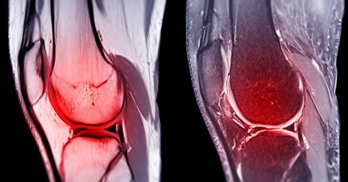
Arthritis and Joint Conditions
Joint conditions like osteoarthritis or rheumatoid arthritis can cause pain, swelling, and stiffness in the arm and hand. MRI can provide detailed images of the joints, showing the degree of cartilage damage, inflammation, or bone degeneration. In some cases, ganglion cysts, which are fluid-filled lumps often linked to joint or tendon issues, can also be identified and evaluated using MRI.
Why MRI Is Essential for Accurate Diagnosis
The precision of MRI makes it a must-have tool for diagnosing arm and hand issues. The detailed images it produces help doctors see exactly what’s going on inside your body, particularly when it comes to soft tissues like tendons, muscles, and nerves. This clarity is essential for identifying subtle injuries or conditions that other imaging techniques might miss.
For example, minor tendon tears or nerve entrapments might not show up on an X-ray, but an MRI can provide the detail needed to pinpoint the problem. This makes MRI particularly valuable when planning treatments or surgeries, as it gives doctors a clear understanding of what’s causing the pain.
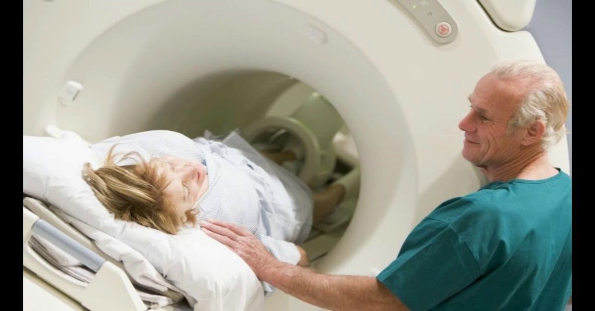
MRI for Post-Treatment and Surgical Follow-Up
Once treatment begins, whether it’s through physical therapy or surgery, MRI continues to play an important role. Doctors often use MRI to monitor healing progress. For instance, after a tendon repair surgery, an MRI can show whether the tendon is healing properly and if any complications, like scar tissue, are developing. This follow-up imaging ensures that everything is on track and that any issues can be addressed early before they become bigger problems.
When Should You Consider an MRI for Arm or Hand Pain?
So when should you consider getting an MRI for your arm or hand? If you’ve been dealing with pain for a while and traditional methods like X-rays or physical exams haven’t provided a clear answer, it might be time to look deeper with an MRI. This is especially true if you’re experiencing nerve-related symptoms like numbness, tingling, or weakness, which could indicate nerve compression or entrapment.
Chronic pain, persistent swelling, or suspected soft tissue damage that hasn’t been confirmed with other tests are also good reasons to consider an MRI. By getting a clear, detailed look inside your arm or hand, you and your doctor can make more informed decisions about your treatment plan.
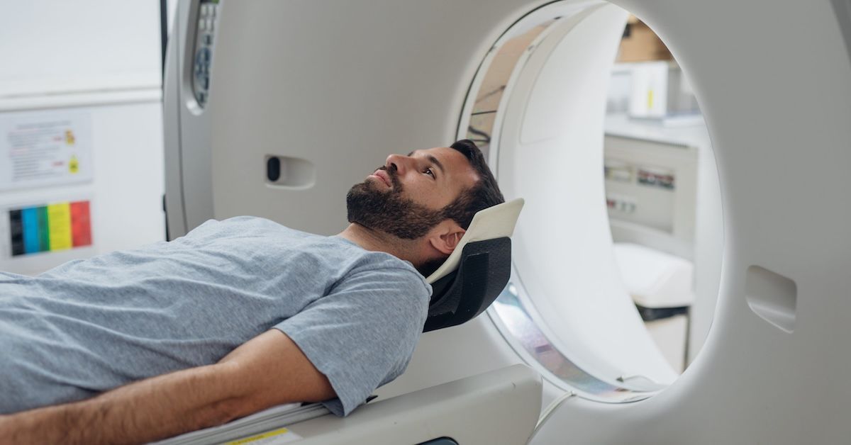
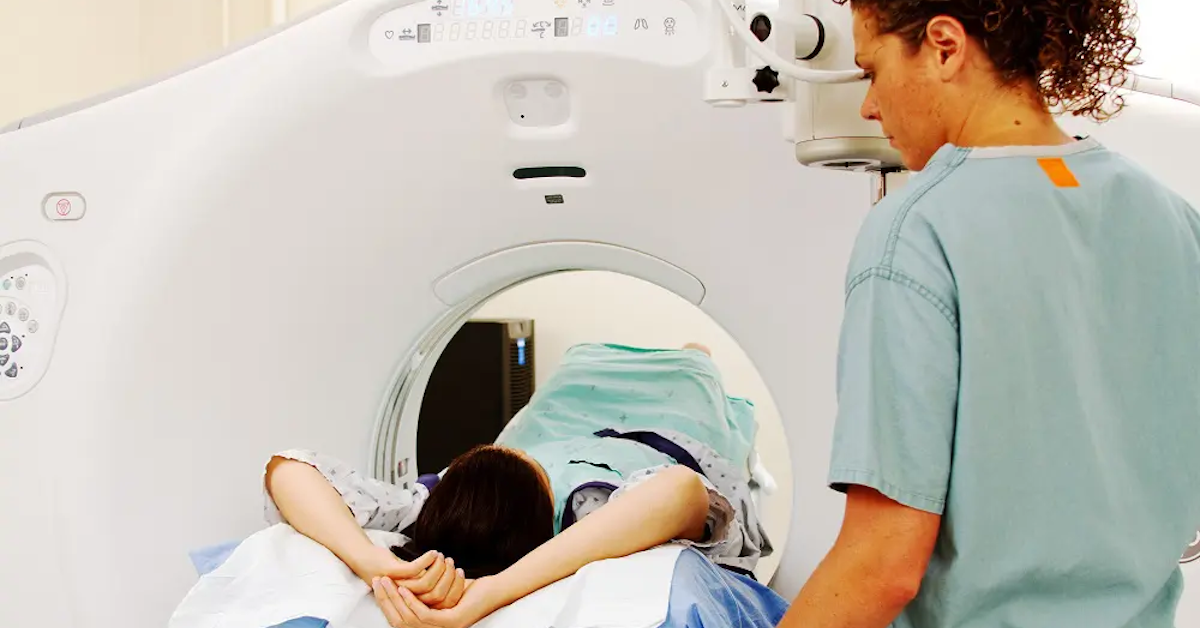
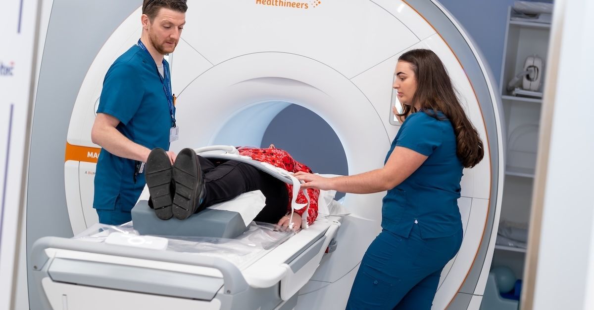
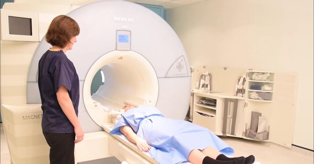
What to Expect During an MRI Scan for the Arm and Hand
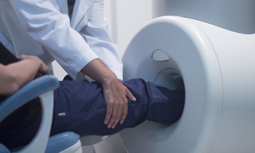
The MRI process is straightforward. You’ll likely be asked to lie down on a table that slides into the MRI machine. For most arm and hand scans, you’ll only need to have your arm positioned inside the machine, leaving the rest of your body outside—so no need to worry about feeling claustrophobic.
The machine will make some noise as it takes images, but you’ll be given earplugs or headphones to help block out the sound. The scan usually takes 30 minutes to an hour, and you’ll need to lie still during this time to ensure clear images. Sometimes, contrast dye is used to enhance the images, but this is typically safe and painless.
Advancements in MRI Technology for Arm and Hand Diagnostics
MRI technology is always evolving, with new advancements making scans faster and clearer. High-resolution imaging now provides more detailed pictures, while open MRI systems are helping patients who may feel uneasy in traditional machines. For some conditions, dynamic MRI—where images are taken while the patient moves their hand or arm—can offer even more insight into problems that only occur during specific motions.
Conclusion
MRI is a powerful tool that offers unmatched detail when diagnosing arm and hand conditions. Whether you’re dealing with a soft tissue injury, a nerve issue, or a bone condition, MRI provides the clarity needed to pinpoint the problem and guide treatment.
At Upright MRI of Deerfield, we specialize in providing high-quality MRI services that give patients the answers they need to get back to feeling their best. If you’re experiencing persistent pain or have been referred for an MRI, we’re here to help make the process as smooth and comfortable as possible.
SHARE THIS POST:
Leave a Comment:

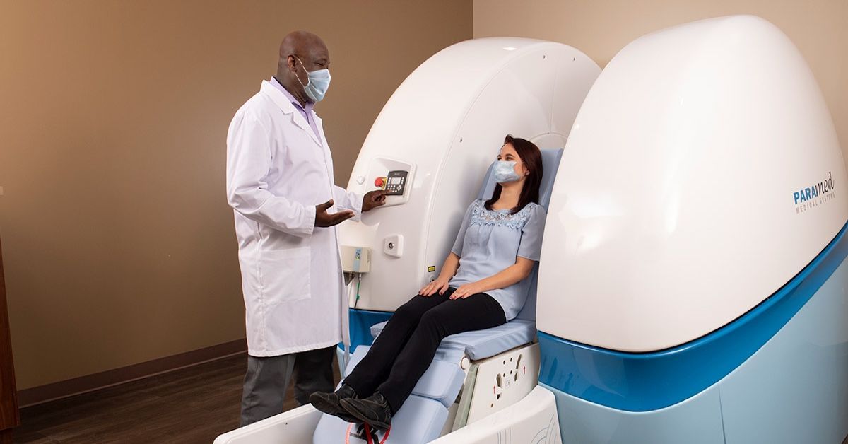
The World's Most Patient-Friendly MRI. A comfortable, stress-free, and completely reliable MRI scan. We offer patients an open, upright, standup MRI experience that helps those who are claustrophobic and stress being in a confined area. Upright MRI of Deerfield is recognized as the world leader in open MRI innovation,
Our Recent Post
READ PATIENT TESTIMONIALS
Upright MRI of Deerfield.
Susan D.,
Highland Park, 39
I am going to tell everyone about your office! This was a great experience after I panicked in other MRI machines and had to leave. Thank you so much.

Judith B.,
Milwaukee, 61
I suffer from vertigo and other MRIs do not work. This was wonderful…absolutely NO discomfort at all. The MRI was so fast…I wanted to stay and watch the movie! Mumtaz was great. His humor really put me at ease. I’ve already recommended Upright MRI to friends.
Delores P.,
Glencoe, 55
Everything is so nice and professional with your place. I have been there a couple of times. My husband and I would not go anywhere else.


