
457 Lake Cook Road (Deerfield Park Plaza)
Deerfield, IL 60015
Email Us:
Email us directly [+]
Fax: (847) 291-9362
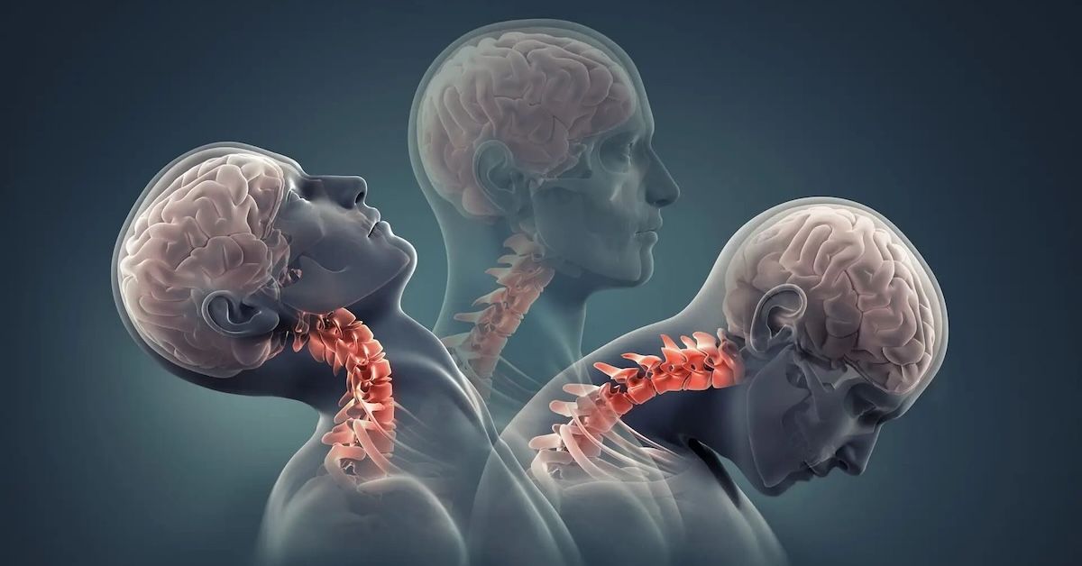
In-Depth Look at MRI Knee Scans: Diagnosing Knee Injuries
Regarding musculoskeletal health, the knee is essential to our daily activities. Our knees take the brunt of it all, whether we're jogging, walking, or just standing. But a variety of ailments and traumas can affect this important joint, severely impairing our movement and quality of life. This is where state-of-the-art technology is useful. The MRI (Magnetic Resonance Imaging) is one such diagnostic wonder. We'll explore the complexities of MRI knee scans in this extensive tutorial, highlighting their importance for accurate diagnosis and treatment planning.
How MRI Works
A miracle of contemporary medicine, magnetic resonance imaging (MRI) creates finely detailed pictures of the body's interior components by combining radio waves and magnetic fields. Nuclear magnetic resonance, which entails aligning hydrogen atoms within the body and then perturbing them with radio waves, is the basis for how it works. These atoms produce signals that are transformed into high-resolution pictures when they return to their original condition. This makes it possible to see the knee joint in great detail and without any kind of surgery.
Advantages of MRI Knee Scans
MRI knee scans provide several benefits. Compared to many other imaging techniques, they enable for a degree of detail that is unmatched in resolution. In addition, MRI scans don't require any intrusive treatments like surgery or injections. They are therefore a safer choice, especially for those who might be uncomfortable with intrusive procedures. Furthermore, MRI offers a thorough understanding of the problem at hand by detecting a broad spectrum of knee injuries and disorders, from ligament tears to bone fractures.
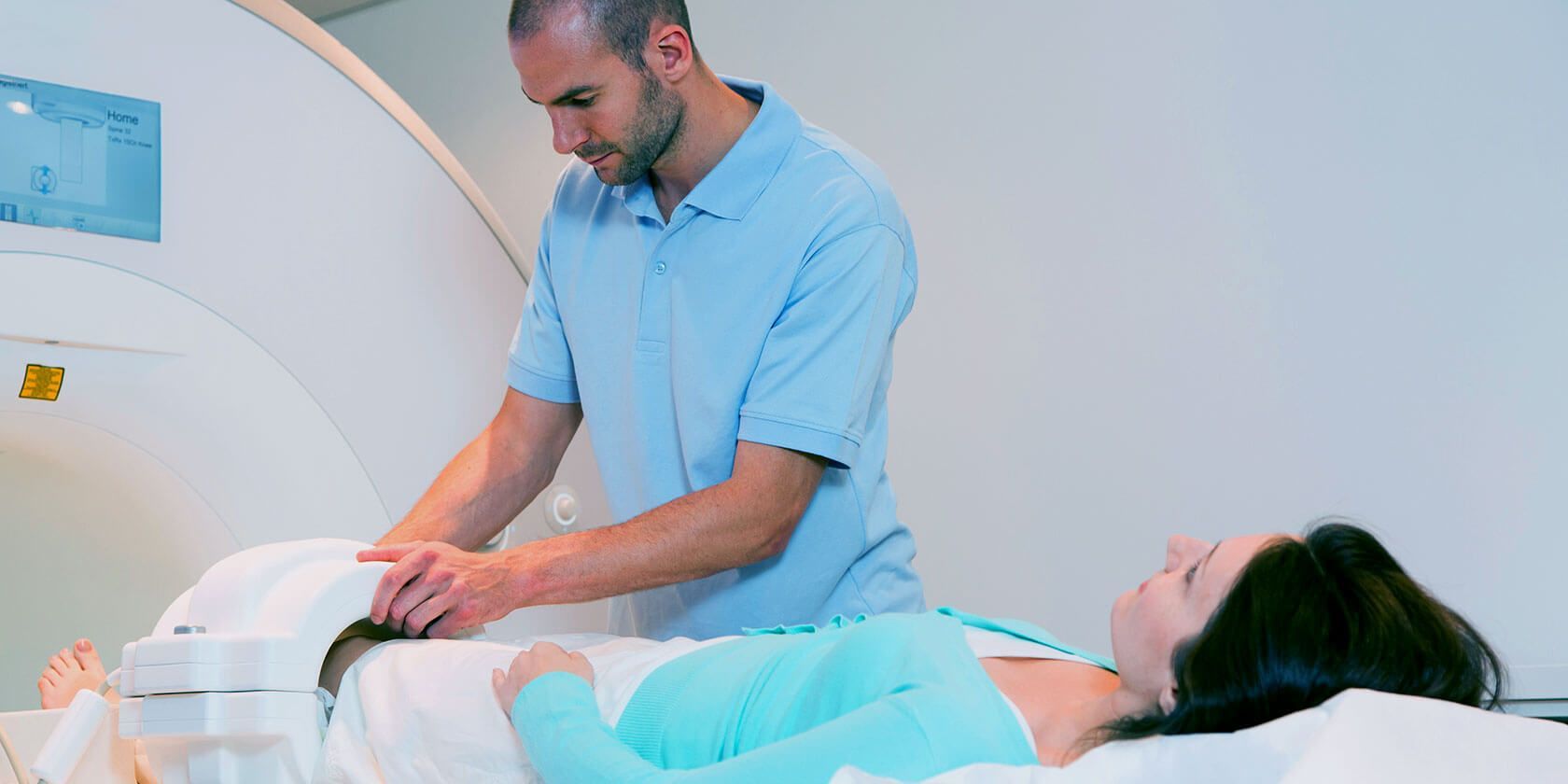
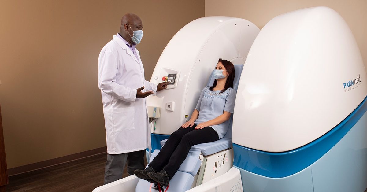
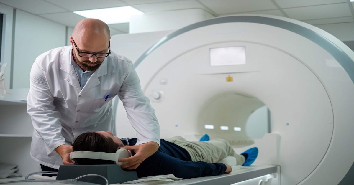
Preparation and Procedure for a Knee MRI
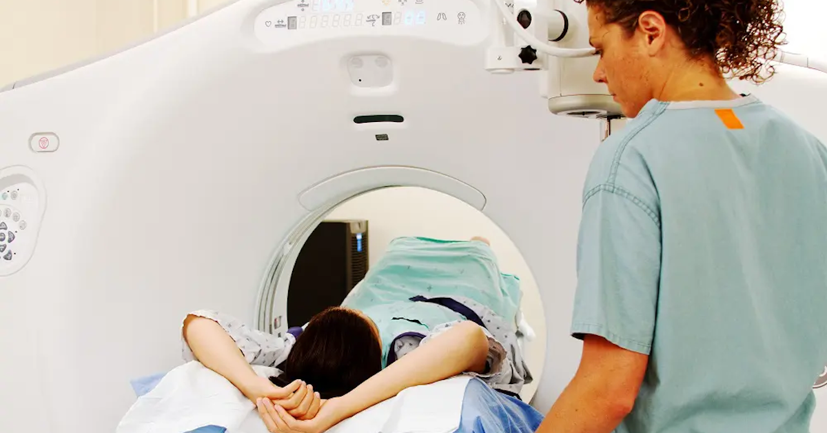
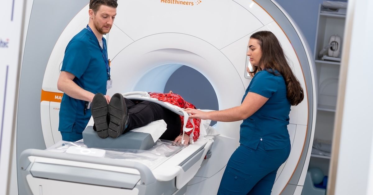
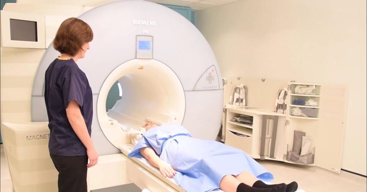
There are a few important things to remember to prepare for a knee MRI appointment. Usually, doctors advise patients to dress comfortably and without metal. Any metal implants or gadgets in your body must be disclosed to the technician, since they may cause interference during the imaging procedure. The MRI equipment will make a number of clicks and hums once it is in a comfortable position and starting taking comprehensive pictures of your knee. The process is painless, but in order to guarantee that the photographs are clear, you must remain still.
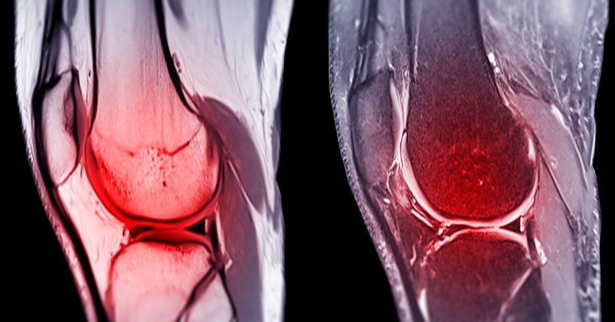
Types of Knee Injuries Detectable with MRI
An extensive range of injuries and disorders may be detected by MRI knee scans, making them highly flexible. These include the typical ligament tears seen in sports and those leading active lives, such as ACL, MCL, PCL, and LCL injuries. Meniscal tears are another common condition that may be detected by MRI and are marked by discomfort and reduced motion. MRI scans can also clearly show signs of tendonitis, rips in the tendon, osteoarthritis, and damage to the cartilage. This sophisticated imaging equipment may even effectively identify contusions and bone fractures.
Interpreting MRI Results
A qualified radiologist with experience in musculoskeletal imaging is needed to interpret MRI data. They will carefully examine the photos, looking for any irregularities or indications of damage. A thorough report is given to patients, sometimes supplemented by illustrations and annotations for easier understanding. For patients looking for clarification on the health of their knees, it is important to comprehend the terminology employed in these reports.
Complementary Techniques: When to Consider Additional Imaging
Even while MRI is an effective diagnostic tool, there are some situations in which other methods may be more appropriate. While ultrasounds are good at seeing soft tissues like tendons and ligaments, X-rays are useful for determining bone density and identifying fractures. In-depth cross-sectional pictures are provided by CT scans, which are very helpful for complicated fractures. To achieve a thorough examination, a variety of imaging techniques may be used, depending on the kind of injury or ailment.
Future Trends in Knee Imaging
Technology is always evolving, and medical imaging is no exception. The field of knee diagnostics might be revolutionized by emerging technology. The goal of developments in open and high-field MRI systems is to improve patient accessibility and comfort. Furthermore, improvements in contrast materials and imaging techniques should make it possible to see knee structures with even more detail. These developments for the future portend favorably for better treatment results and patient care.
Conclusion
It is impossible to overestimate the significance of an accurate diagnosis in the field of musculoskeletal health. MRI knee scans are a ray of hope because they provide unmatched understanding of the complexity of knee ailments and injuries. Their non-invasive nature and high resolution capabilities have made them indispensable in contemporary orthopedics. Seeking expert medical counsel and thinking about an MRI scan can help people with knee problems get back on the road to recovery. Our mission at Upright MRI of Deerfield is to assist you on your path to the best possible musculoskeletal health by offering state-of-the-art imaging solutions.
SHARE THIS POST:
Leave a Comment:
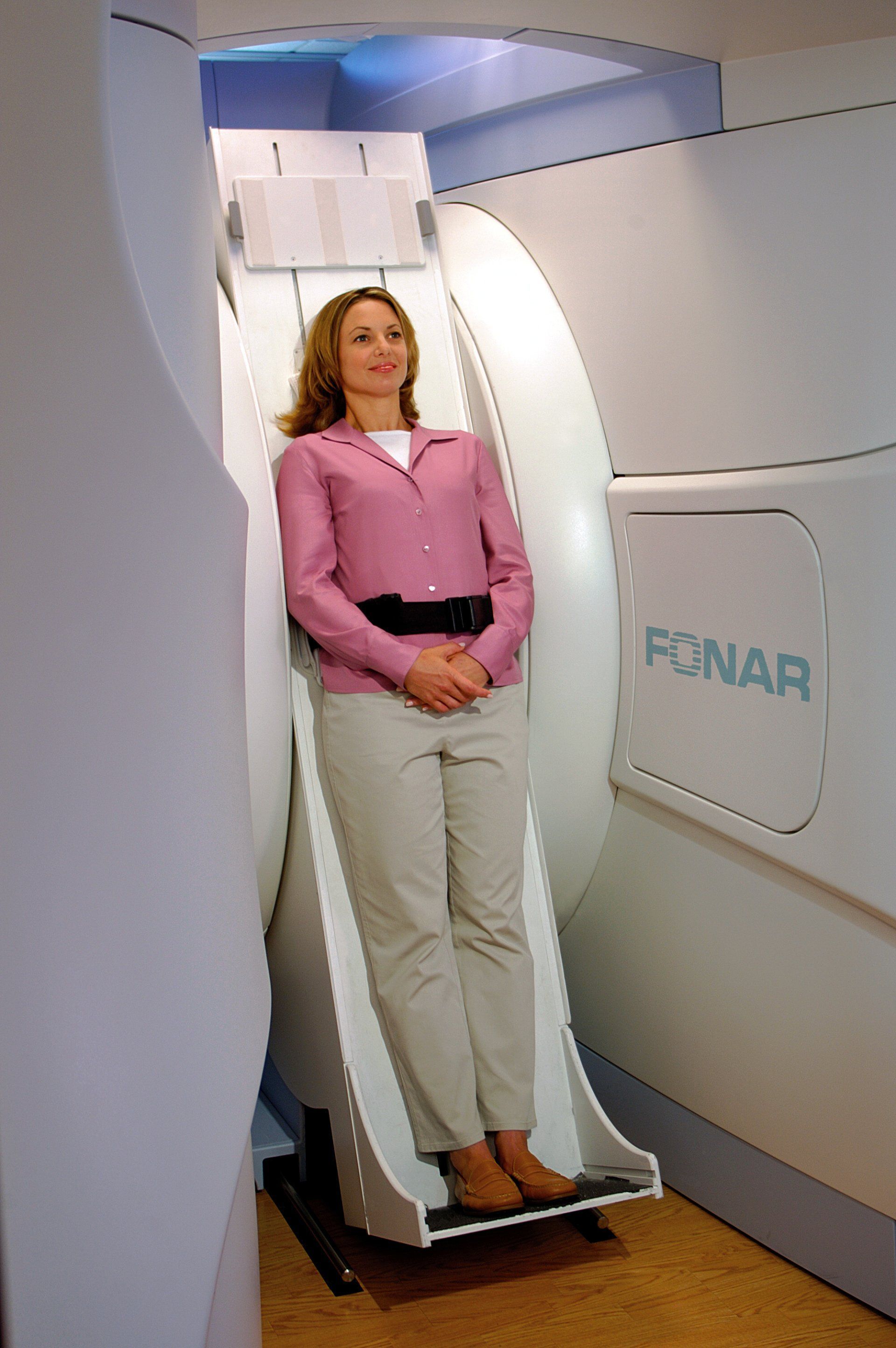
The World's Most Patient-Friendly MRI. A comfortable, stress-free, and completely reliable MRI scan. We offer patients an open, upright, standup MRI experience that helps those who are claustrophobic and stress being in a confined area. Upright MRI of Deerfield is recognized as the world leader in open MRI innovation,
Our Recent Post


READ PATIENT TESTIMONIALS
Upright MRI of Deerfield.
Susan D.,
Highland Park, 39
I am going to tell everyone about your office! This was a great experience after I panicked in other MRI machines and had to leave. Thank you so much.

Judith B.,
Milwaukee, 61
I suffer from vertigo and other MRIs do not work. This was wonderful…absolutely NO discomfort at all. The MRI was so fast…I wanted to stay and watch the movie! Mumtaz was great. His humor really put me at ease. I’ve already recommended Upright MRI to friends.
Delores P.,
Glencoe, 55
Everything is so nice and professional with your place. I have been there a couple of times. My husband and I would not go anywhere else.

Follow UpRight MRI of Deerfield on Facebook
To see our latest news, updates or to get to know us more, we welcome you to follow along our journey in Facebook.


CONTACT DETAILS
Phone: (847) 291-9321
Address: 457 Lake Cook Road (Deerfield Park Plaza) Deerfield, IL 60015
Email: info@uprightmrideerfield.com
Business Hours
- Mon - Thu
- -
- Friday
- -
- Saturday
- -
- Sunday
- Closed