
457 Lake Cook Road (Deerfield Park Plaza)
Deerfield, IL 60015
Email Us:
Email us directly [+]
Fax: (847) 291-9362
What Conditions Can an Upright MRI Detect That a Standard MRI Cannot?
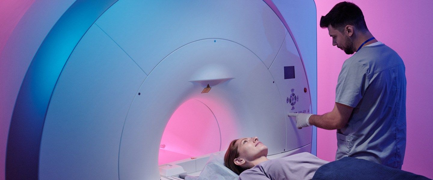
In the realm of medical imaging, Magnetic Resonance Imaging (MRI) has long been a stalwart in providing detailed insights into the human body's inner workings. However, the conventional, supine-position MRI, while immensely valuable, has its limitations. Imagine a diagnostic tool that could transcend these limitations, offering a more comprehensive understanding of certain conditions. Enter the Upright MRI – a revolutionary approach that challenges the constraints of traditional imaging.
Understanding Standard MRI Limitations
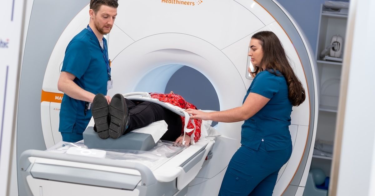
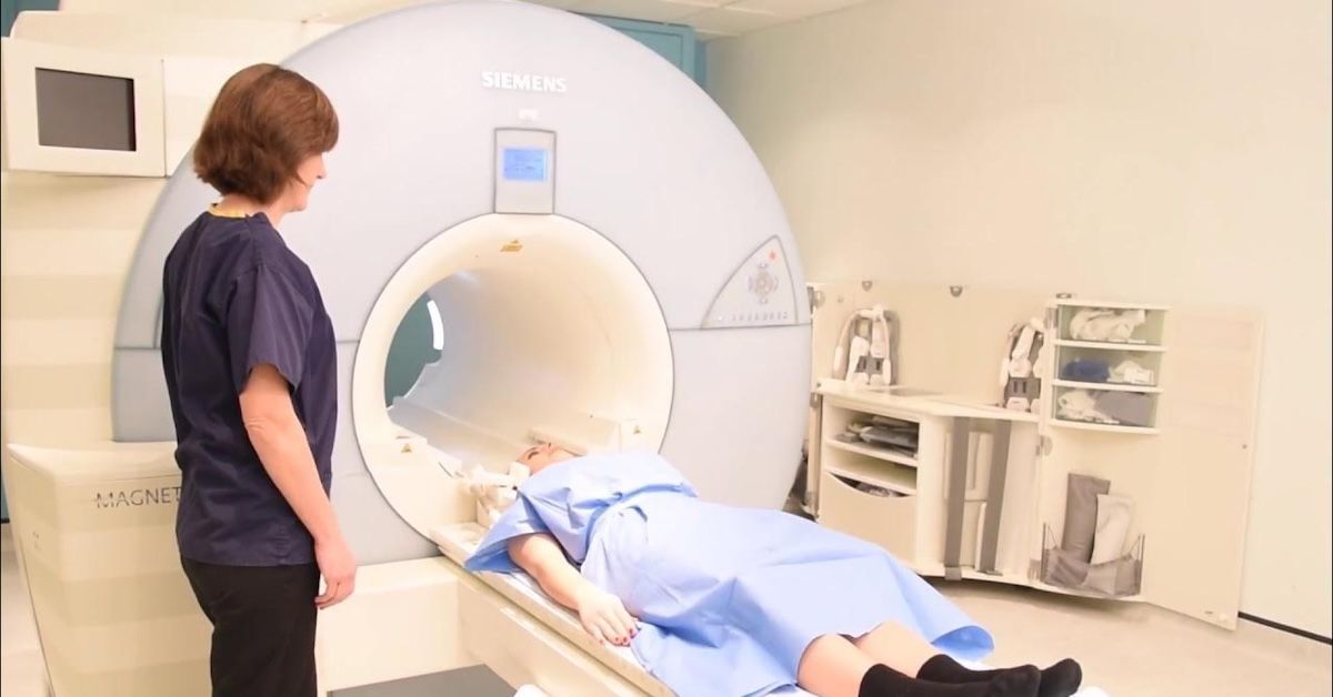
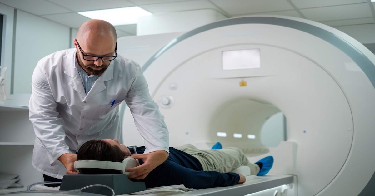
Traditional MRI procedures typically require patients to lie down in a supine position, limiting the range of conditions that can be effectively detected. The static nature of these scans often fails to capture the dynamic changes that occur when the body is in its natural weight-bearing state. Consequently, certain conditions that manifest or exacerbate under load may be overlooked, leaving patients with incomplete diagnoses and healthcare providers with incomplete pictures.
The Evolution of Upright MRI
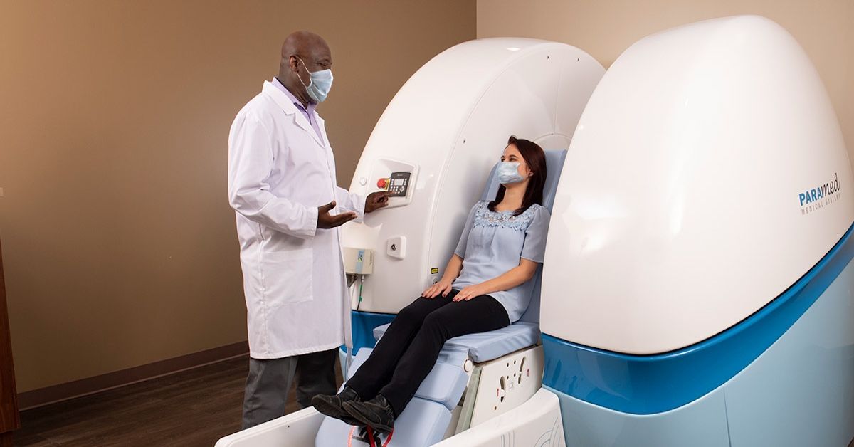
In response to the constraints of standard MRI, the Upright MRI emerged as a game-changer in diagnostic imaging. The fundamental shift lies in patient positioning. Unlike traditional MRI, where patients are confined to a horizontal position, upright MRI allows imaging in weight-bearing positions, providing a more realistic representation of the body's dynamics. This innovative approach not only enhances diagnostic accuracy but also broadens the scope of conditions that can be effectively identified.
Conditions Detected by Upright MRI
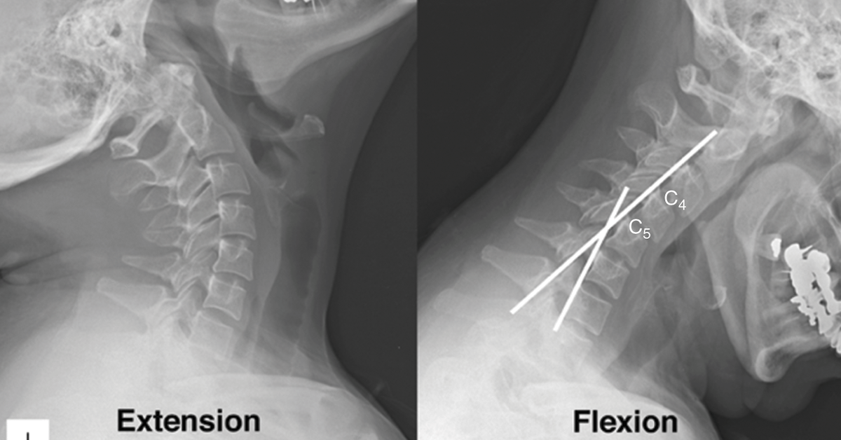
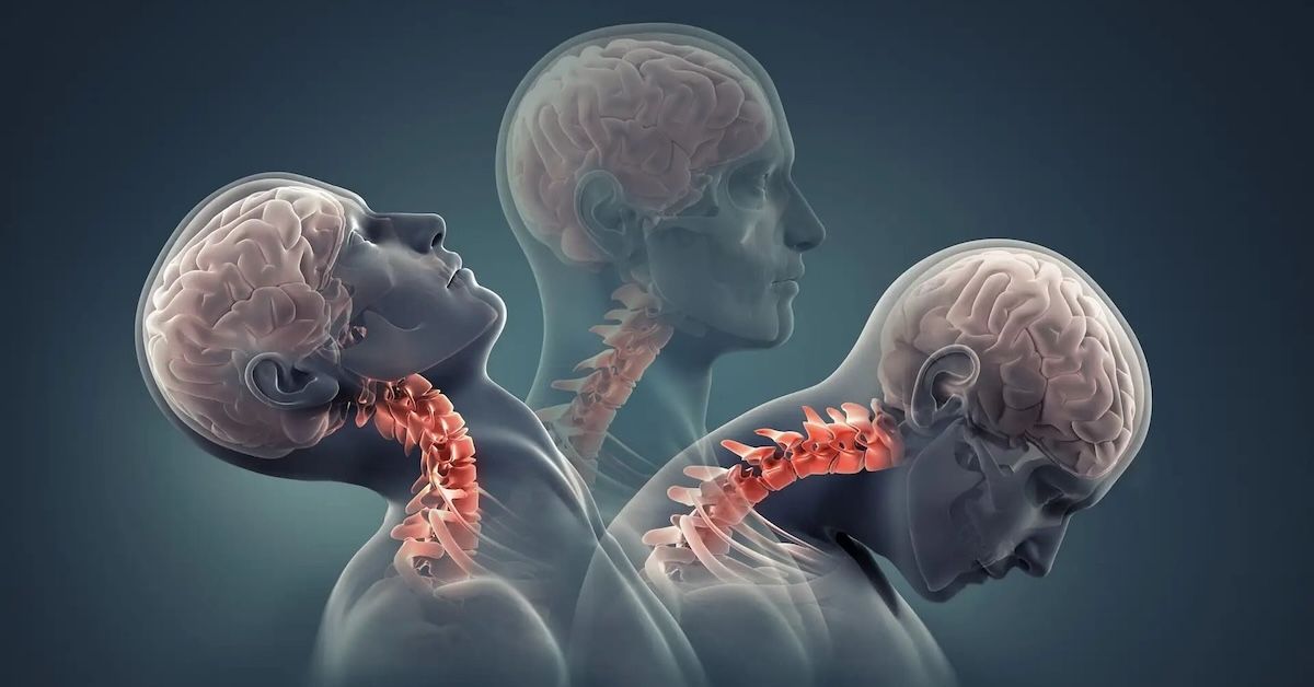
The versatility of upright MRI becomes particularly evident when considering conditions affecting the spine, joints, and neurological system. In the realm of spinal health, upright MRI excels in visualizing dynamic changes that occur when the spine bears the body's weight. Conditions such as disc herniation, spinal stenosis, and scoliosis, which may go unnoticed in standard MRI, become discernible in the upright setting.
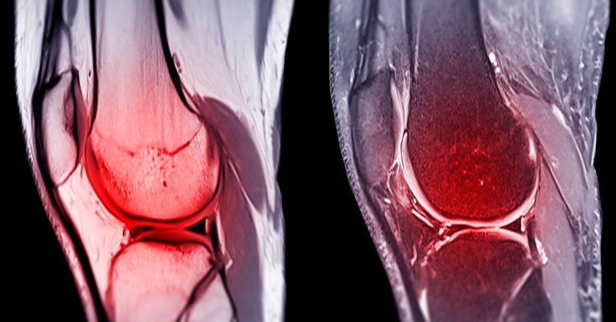
Joint-related issues, often elusive in traditional scans, also come to light with upright MRI. Whether it's assessing the temporomandibular joint (TMJ) or scrutinizing knee and hip conditions, the weight-bearing aspect provides a more comprehensive understanding of joint function. This added dimension is crucial for accurate diagnosis and subsequent treatment planning.
Moving beyond the musculoskeletal system, upright MRI unveils its prowess in detecting neurological disorders. Conditions like Chiari malformation, characterized by the displacement of the cerebellum, are better visualized when patients are upright. Similarly, intracranial hypotension, a condition often associated with low cerebrospinal fluid pressure, can be more precisely diagnosed through positional imaging.
Advantages of Upright MRI in Specific Cases
The advantages of upright MRI extend beyond merely detecting conditions; they lie in the depth of insight it provides in specific cases. For spinal conditions, the ability to visualize dynamic changes during weight-bearing enables a more thorough evaluation. This is particularly crucial in identifying spinal instability, a factor that might be underestimated in conventional MRI scans.
In joint-related issues, upright MRI offers a unique advantage by allowing clinicians to evaluate joint function in a natural, weight-bearing state. This proves invaluable in discerning subtle abnormalities that might be missed in standard MRI scans, ultimately guiding orthopedic interventions with greater precision.
When it comes to neurological disorders, the positional imaging offered by upright MRI plays a pivotal role. Chiari malformation, for instance, may only become apparent when the cerebellum undergoes displacement under the influence of gravity. Upright MRI's ability to capture these nuances contributes significantly to accurate diagnoses and subsequent treatment strategies.
Patient Experience and Safety
Beyond its diagnostic capabilities, the Upright MRI brings added comfort to patients. The experience of undergoing imaging in a more natural, weight-bearing posture can be more tolerable, particularly for those with conditions that worsen or become apparent in specific positions. Additionally, safety concerns are addressed with protocols comparable to standard MRI, ensuring patients' well-being throughout the procedure.
Conclusion
In the ever-evolving landscape of medical diagnostics, the Upright MRI stands as a beacon of progress, offering a more comprehensive and nuanced approach to imaging. As we delve into the intricacies of spinal, joint, and neurological conditions, the significance of upright MRI becomes apparent. It is not merely a tool for detection; it is a gateway to a deeper understanding of the body in motion.
As we navigate the future of diagnostic imaging, it is imperative to consider the expanded possibilities that upright MRI brings to the table. For those seeking a more holistic evaluation of certain conditions, this innovative technology is poised to redefine the diagnostic landscape. In the quest for precision and depth, Upright MRI emerges as a formidable ally in the hands of healthcare providers, unraveling mysteries that traditional imaging might overlook.
This exploration into the capabilities of upright MRI is brought to you by Upright MRI, a pioneering force in diagnostic imaging. Committed to advancing patient care through cutting-edge technology, Upright MRI is at the forefront of revolutionizing how we perceive and address various health conditions. Harnessing the power of upright imaging, we strive to empower healthcare providers with the tools they need to make more informed decisions and, in turn, improve patient outcomes.
SHARE THIS POST:
Leave a Comment:
The World's Most Patient-Friendly MRI. A comfortable, stress-free, and completely reliable MRI scan. We offer patients an open, upright, standup MRI experience that helps those who are claustrophobic and stress being in a confined area. Upright MRI of Deerfield is recognized as the world leader in open MRI innovation,
Our Recent Post


READ PATIENT TESTIMONIALS
Upright MRI of Deerfield.
Susan D.,
Highland Park, 39
I am going to tell everyone about your office! This was a great experience after I panicked in other MRI machines and had to leave. Thank you so much.

Judith B.,
Milwaukee, 61
I suffer from vertigo and other MRIs do not work. This was wonderful…absolutely NO discomfort at all. The MRI was so fast…I wanted to stay and watch the movie! Mumtaz was great. His humor really put me at ease. I’ve already recommended Upright MRI to friends.
Delores P.,
Glencoe, 55
Everything is so nice and professional with your place. I have been there a couple of times. My husband and I would not go anywhere else.

Follow UpRight MRI of Deerfield on Facebook
To see our latest news, updates or to get to know us more, we welcome you to follow along our journey in Facebook.


CONTACT DETAILS
Phone: (847) 291-9321
Address: 457 Lake Cook Road (Deerfield Park Plaza) Deerfield, IL 60015
Email: info@uprightmrideerfield.com
Business Hours
- Mon - Thu
- -
- Friday
- -
- Saturday
- -
- Sunday
- Closed