
457 Lake Cook Road (Deerfield Park Plaza)
Deerfield, IL 60015
Email Us:
Email us directly [+]
Fax: (847) 291-9362
What Insights Can MRI Imaging Offer for Shoulder Injuries?
Shoulder pain is more than just an inconvenience. It can impact your daily routine, making simple tasks like reaching for a shelf or lifting groceries difficult. Accurate diagnosis is key to effective treatment, and that's where MRI imaging shines. MRI scans provide detailed images of the shoulder, uncovering issues that other imaging techniques might miss. Let's delve into how MRI imaging can offer crucial insights into shoulder injuries.
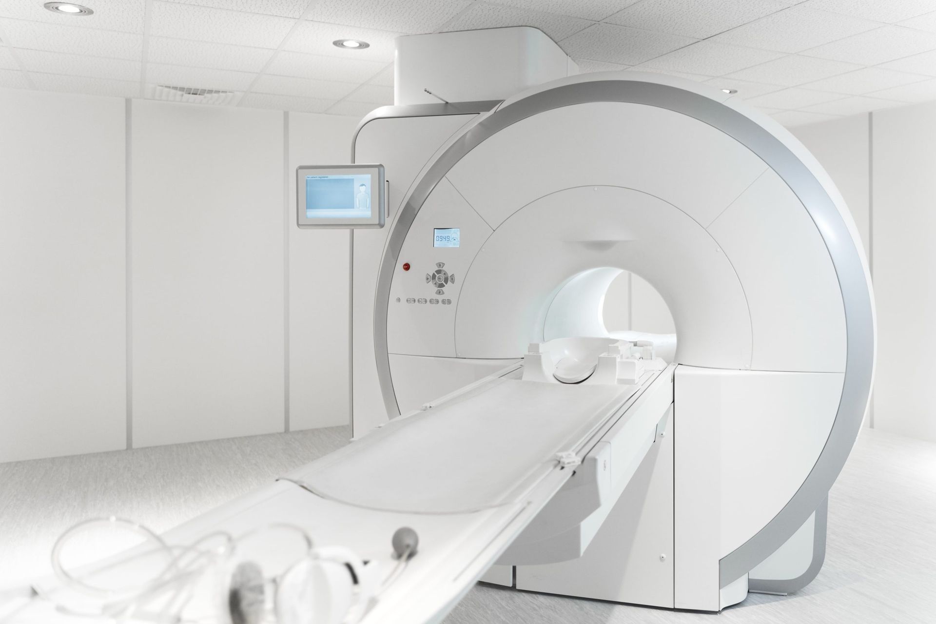
Understanding MRI Imaging
MRI, or Magnetic Resonance Imaging, is a powerful tool in medical diagnostics. It uses strong magnets and radio waves to create detailed images of the body's internal structures. Unlike X-rays, which are great for seeing bones, MRIs are excellent at visualizing soft tissues like muscles, tendons, and ligaments. This makes MRI particularly useful for diagnosing shoulder injuries, where these tissues are often involved.
Common Shoulder Injuries
The shoulder is a complex joint with a lot of moving parts, making it prone to various injuries. Here’s a quick look at some common ones:
- Rotator Cuff Tears: These are tears in the muscles and tendons that hold the shoulder joint in place.
- Labral Tears: Damage to the cartilage that surrounds the shoulder socket.
- Tendinitis: Inflammation of the tendons, often due to overuse.
- Bursitis: Inflammation of the bursa, a fluid-filled sac that reduces friction in the joint.
- Shoulder Impingement: Occurs when shoulder blade rubs against the rotator cuff.
- Dislocations and Fractures: Injuries to the bones of the shoulder, such as the humerus, scapula, or clavicle.
Insights MRI Imaging Offers for Shoulder Injuries
MRI imaging offers a range of benefits in diagnosing shoulder injuries, providing a level of detail that can significantly impact treatment plans.
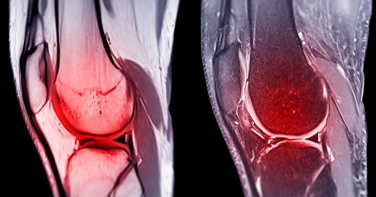
Detailed Visualization of Soft Tissues
One of the standout features of MRI is its ability to clearly show soft tissues. This means doctors can see muscles, tendons, ligaments, and cartilage in great detail. For shoulder injuries, this level of clarity is crucial. It helps in identifying issues like small tears in the rotator cuff or inflammation in the tendons that other imaging methods might miss.
Detection of Tears and Strains
MRI is particularly good at detecting tears and strains. Whether it’s a partial tear in the rotator cuff or a complete tear in the labrum, MRI can pinpoint the exact location and severity. This precision is vital for determining the best course of action, whether it's physical therapy, injections, or surgery.
Assessment of Inflammation and Swelling
Inflammation and swelling are common in many shoulder injuries. MRI can detect these conditions, showing areas of increased fluid or thickened tissues. Identifying inflammation helps doctors understand the extent of an injury and can guide treatments like anti-inflammatory medications or cortisone injections.
Evaluation of Structural Abnormalities
Structural issues, such as bone spurs or cartilage damage, can also be seen with MRI. These abnormalities might contribute to conditions like shoulder impingement, where the bones pinch the rotator cuff. By revealing these problems, MRI helps doctors plan treatments that address the root cause, not just the symptoms.
Diagnosis of Complex Conditions
Some shoulder injuries are more complex and harder to diagnose. Conditions like labral tears and SLAP lesions (a specific type of labral tear) require detailed imaging to be properly identified. MRI’s high-resolution images can show these intricate details, leading to more accurate diagnoses and better treatment outcomes.
The MRI Procedure for Shoulder Injuries
Understanding what happens during an MRI can make the process less daunting.
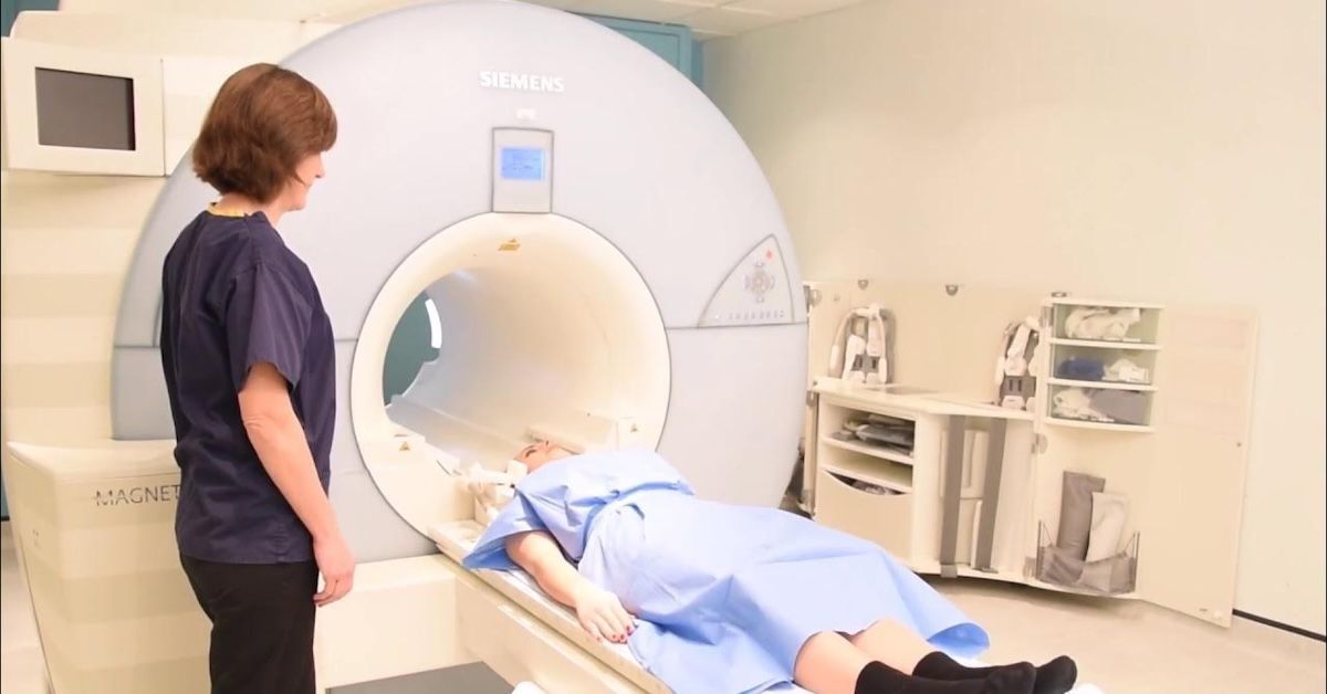
Preparation for the Scan
Before your MRI, you’ll need to remove any metal objects and change into a gown. It’s also important to inform the technician if you have any metal implants or devices in your body, as these can interfere with the magnetic field.
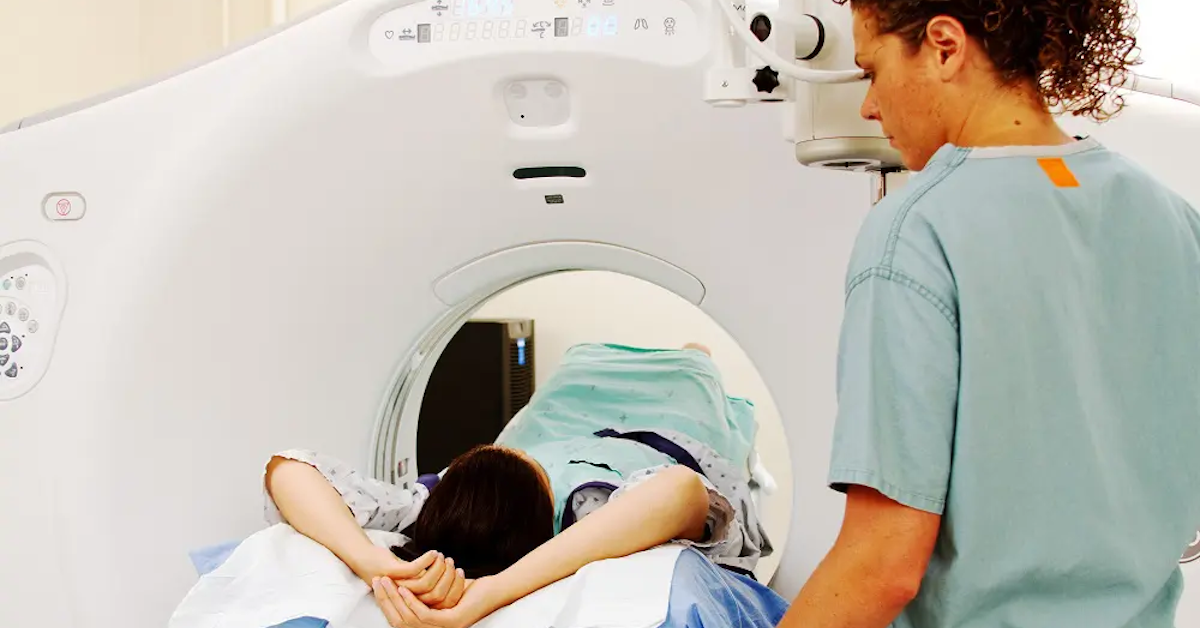
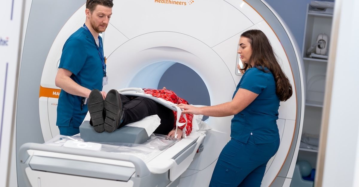
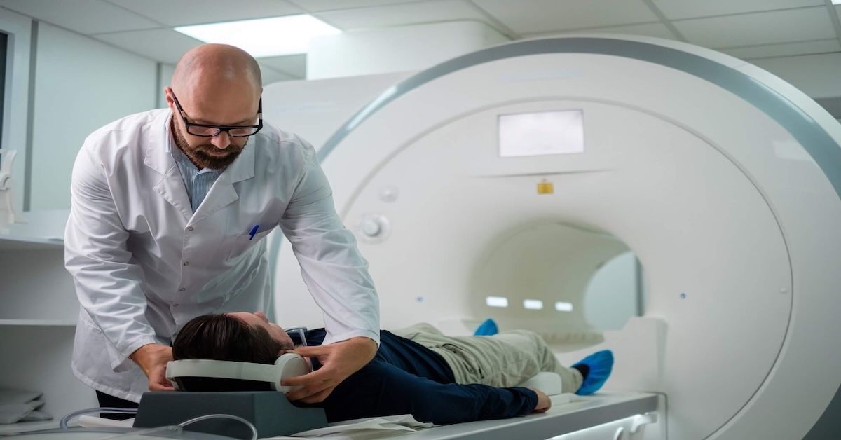
What to Expect During the Scan
During the scan, you’ll lie down on a table that slides into the MRI machine. The process is painless, but you’ll need to stay still to ensure clear images. The machine can be loud, making knocking and humming sounds, but you’ll be given earplugs or headphones to reduce the noise. The technician will communicate with you through a microphone to ensure you're comfortable throughout the procedure.
Post-Scan Process
After the scan, you can immediately return to your normal activities. The radiologist will analyze the images and send a report to your doctor. Your doctor will then discuss the findings with you and outline the next steps.
Interpreting MRI Results
Reading MRI results can be complex, but here’s a simple breakdown:
Understanding the Report
An MRI report includes detailed descriptions of what was found during the scan. It’ll mention any tears, inflammation, or structural abnormalities. The report uses medical terms, so don’t hesitate to ask your doctor to explain anything that’s unclear.
Common Findings in MRI Scans
Typical results might show soft tissue injuries like rotator cuff tears, joint abnormalities, or signs of inflammation. Each finding helps paint a picture of what’s causing your shoulder pain and guides the treatment plan.
Consulting with Specialists
Discussing MRI results with specialists, such as orthopedic surgeons or radiologists, is crucial. They can provide a comprehensive diagnosis and recommend the best course of action based on the MRI findings.
Treatment and Management Based on MRI Insights
Once the MRI has provided a clear diagnosis, your doctor can tailor a treatment plan to your specific needs.
Conservative Treatments
For many shoulder injuries, conservative treatments like physical therapy, medications, and lifestyle modifications can be very effective. Physical therapy helps strengthen the shoulder and improve flexibility, while medications can reduce pain and inflammation.
Surgical Interventions
In cases where conservative treatments aren’t enough, surgery might be necessary. MRI results are critical in planning these procedures. For instance, they help surgeons determine the exact location and extent of a tear, ensuring more precise and effective surgery.
Rehabilitation and Recovery
Recovery doesn’t end with surgery or initial treatment. Post-treatment rehabilitation is crucial for a full recovery. Physical therapy, guided by MRI results, helps ensure that the shoulder heals correctly and regains full function.
Future of MRI Technology in Shoulder Injury Diagnosis
MRI technology continues to evolve, promising even better diagnostic capabilities in the future.
Advancements in MRI Technology
New advancements are making MRI scans faster and more comfortable for patients. Improved imaging techniques provide even clearer and more detailed images, aiding in more accurate diagnoses.
Potential Developments
Future developments in MRI technology may include enhanced imaging methods that offer even more precise views of the shoulder. These advancements could lead to earlier detection of injuries and more targeted treatments, improving patient outcomes.
Conclusion
MRI shoulder scans are an invaluable tool in diagnosing and treating shoulder injuries. They offer detailed images of soft tissues, detect tears and inflammation, and reveal structural abnormalities that other imaging methods might miss. Whether you’re dealing with a sports injury or chronic shoulder pain, an MRI can provide the insights needed for effective treatment. For expert advice and state-of-the-art imaging, contact Upright MRI of Deerfield. We’re here to help you get back to your best self.
SHARE THIS POST:
Leave a Comment:
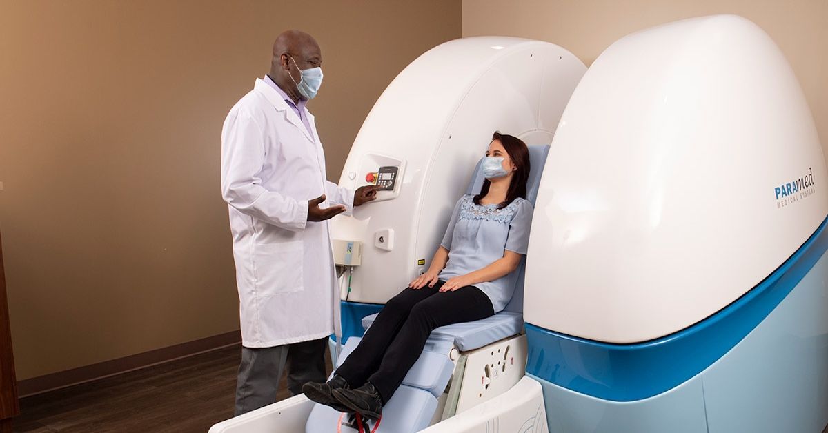
The World's Most Patient-Friendly MRI. A comfortable, stress-free, and completely reliable MRI scan. We offer patients an open, upright, standup MRI experience that helps those who are claustrophobic and stress being in a confined area. Upright MRI of Deerfield is recognized as the world leader in open MRI innovation,
Our Recent Post


READ PATIENT TESTIMONIALS
Upright MRI of Deerfield.
Susan D.,
Highland Park, 39
I am going to tell everyone about your office! This was a great experience after I panicked in other MRI machines and had to leave. Thank you so much.

Judith B.,
Milwaukee, 61
I suffer from vertigo and other MRIs do not work. This was wonderful…absolutely NO discomfort at all. The MRI was so fast…I wanted to stay and watch the movie! Mumtaz was great. His humor really put me at ease. I’ve already recommended Upright MRI to friends.
Delores P.,
Glencoe, 55
Everything is so nice and professional with your place. I have been there a couple of times. My husband and I would not go anywhere else.

Follow UpRight MRI of Deerfield on Facebook
To see our latest news, updates or to get to know us more, we welcome you to follow along our journey in Facebook.


CONTACT DETAILS
Phone: (847) 291-9321
Address: 457 Lake Cook Road (Deerfield Park Plaza) Deerfield, IL 60015
Email: info@uprightmrideerfield.com
Business Hours
- Mon - Thu
- -
- Friday
- -
- Saturday
- -
- Sunday
- Closed