How Can MRI Scans Assist in Diagnosing Cranio-Cervical Instability?
Cranio-cervical instability (CCI) is a condition that affects the junction where the skull meets the cervical spine. It’s a complex issue that can cause a range of symptoms, from chronic neck pain to neurological problems. Diagnosing CCI requires precision, as the condition often involves subtle changes in ligaments, bones, and other structures. MRI (Magnetic Resonance Imaging) has become a vital tool in identifying and understanding CCI, thanks to its ability to provide detailed images without invasive procedures. Here’s how MRI scans can assist in diagnosing cranio-cervical instability and why they’re an invaluable resource for patients and doctors alike.
Understanding Cranio-Cervical Instability
Cranio-cervical instability occurs when the ligaments and structures connecting the skull and cervical spine are weakened or damaged. This instability can lead to abnormal movement, compression of nerves, and strain on the spinal cord.
The causes of CCI are varied. Some people develop it after trauma, like a car accident or sports injury, while others experience it as a complication of connective tissue disorders such as Ehlers-Danlos Syndrome. Degenerative conditions and inflammatory diseases like rheumatoid arthritis can also play a role.
Symptoms of CCI often overlap with other conditions, which can make diagnosis challenging. Patients may experience headaches, neck pain, dizziness, or even neurological issues like tingling in the limbs. Because these symptoms can be vague, imaging tools like MRI are essential for pinpointing the root cause.
Why Imaging Is Crucial for Diagnosing CCI
When a doctor suspects cranio-cervical instability, imaging is often the next step after a physical exam. However, traditional methods like X-rays or CT scans have limitations. X-rays are good for seeing bones but can’t capture the soft tissues or ligaments. CT scans provide more detail but still fall short in assessing the subtle complexities of CCI.
MRI, on the other hand, excels at visualizing both soft tissues and bone structures. It provides a complete picture of the cranio-cervical junction, allowing doctors to assess ligament damage, inflammation, and other critical factors. This level of detail is key in diagnosing CCI accurately and creating a treatment plan tailored to the patient’s needs.
How MRI Detects Cranio-Cervical Instability
The strength of MRI lies in its ability to look beyond the surface. For CCI, MRI focuses on the ligaments that stabilize the cranio-cervical junction, particularly the alar and transverse ligaments. Damage or laxity in these ligaments often underpins the instability.
MRI can also evaluate inflammation or swelling in the area, which may indicate chronic strain or injury. Additionally, MRI scans allow doctors to measure alignment angles, like the basilar angle index (BAI), which helps determine whether the skull and cervical spine are positioned correctly.
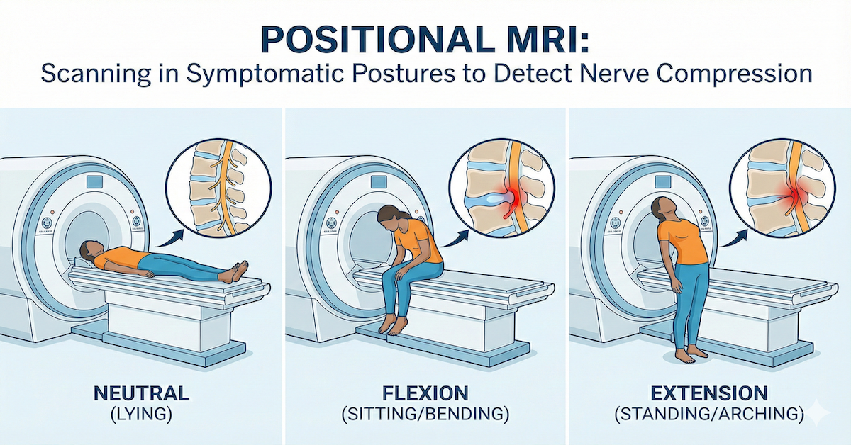
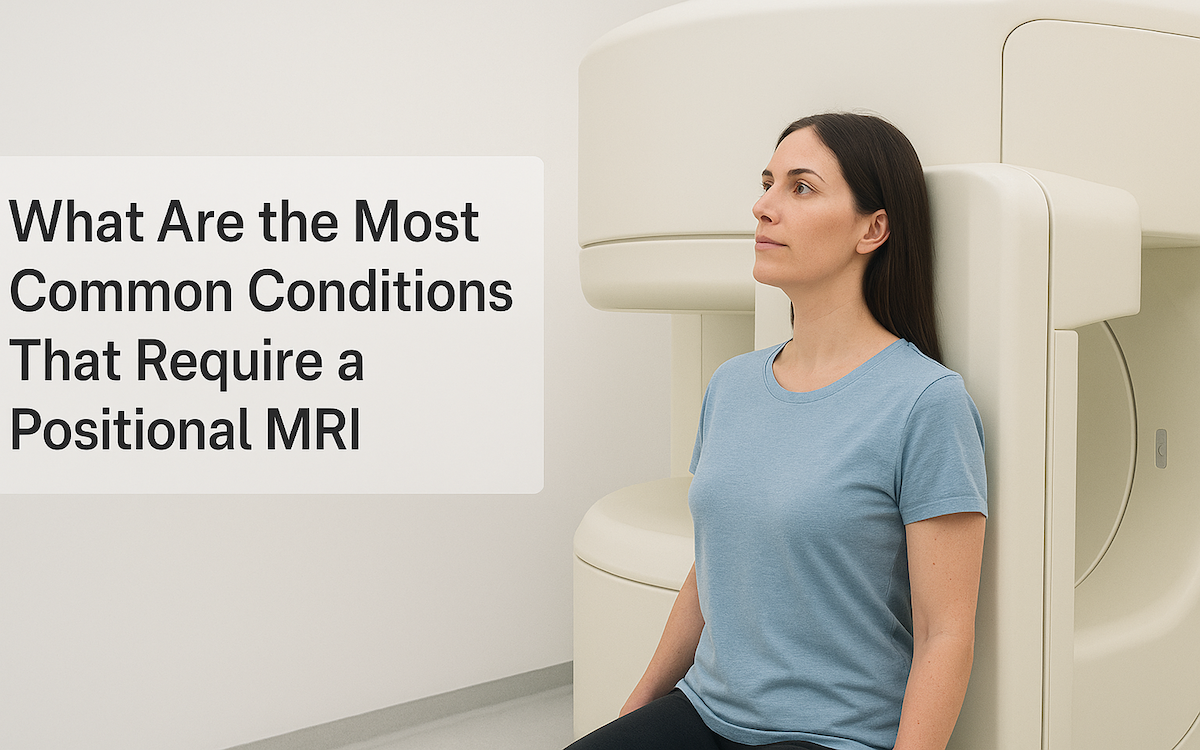
In some cases, dynamic or flexion-extension MRI is used. This technique involves imaging the neck in different positions to assess how the structures move and whether instability occurs during certain motions. This real-time assessment provides critical insights that static imaging methods can’t offer.
Advantages of MRI in Diagnosing CCI
MRI offers several benefits over other imaging techniques. First and foremost, it’s non-invasive. There’s no need for needles or surgical procedures, making it a safer choice for patients. Unlike X-rays or CT scans, MRI doesn’t use radiation, which means it can be repeated if necessary without additional risks.
The high-resolution images produced by MRI allow for an unparalleled view of soft tissues, ligaments, and the spinal cord. This clarity is particularly important in conditions like CCI, where the issue often lies in subtle changes or damage that other imaging tools might miss.
Dynamic MRI further enhances the diagnostic process by showing how the cranio-cervical junction behaves under movement. This provides a clearer picture of how instability impacts the patient’s daily life and helps doctors plan effective treatments.
What to Expect During an MRI for CCI
If you’re scheduled for an MRI to investigate cranio-cervical instability, you may wonder what the procedure entails. Fortunately, it’s a straightforward and painless process.
Before the scan, you’ll be asked to remove any metal objects, as the MRI machine uses strong magnets. Depending on your doctor’s instructions, you might need to fast for a few hours or drink water to stay hydrated.
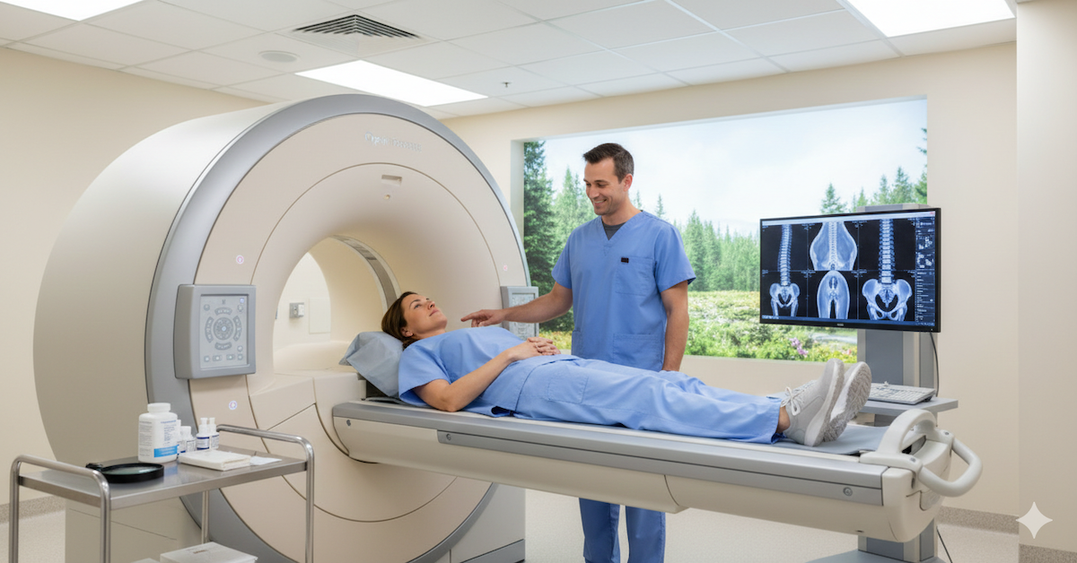
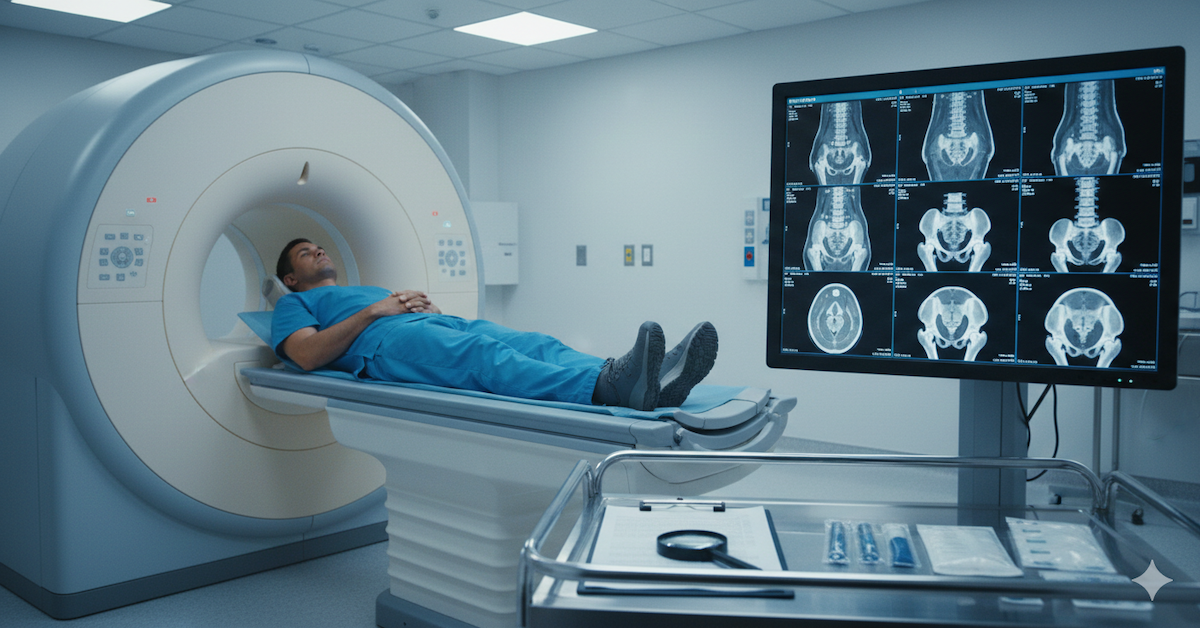
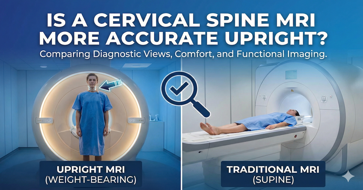
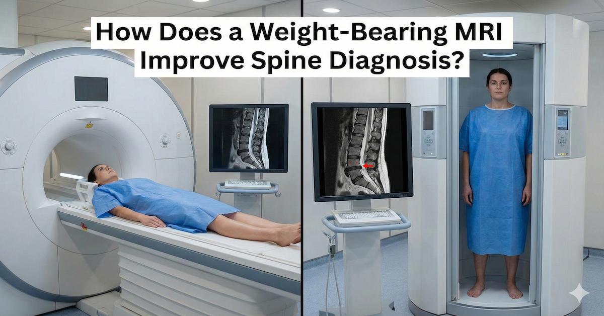
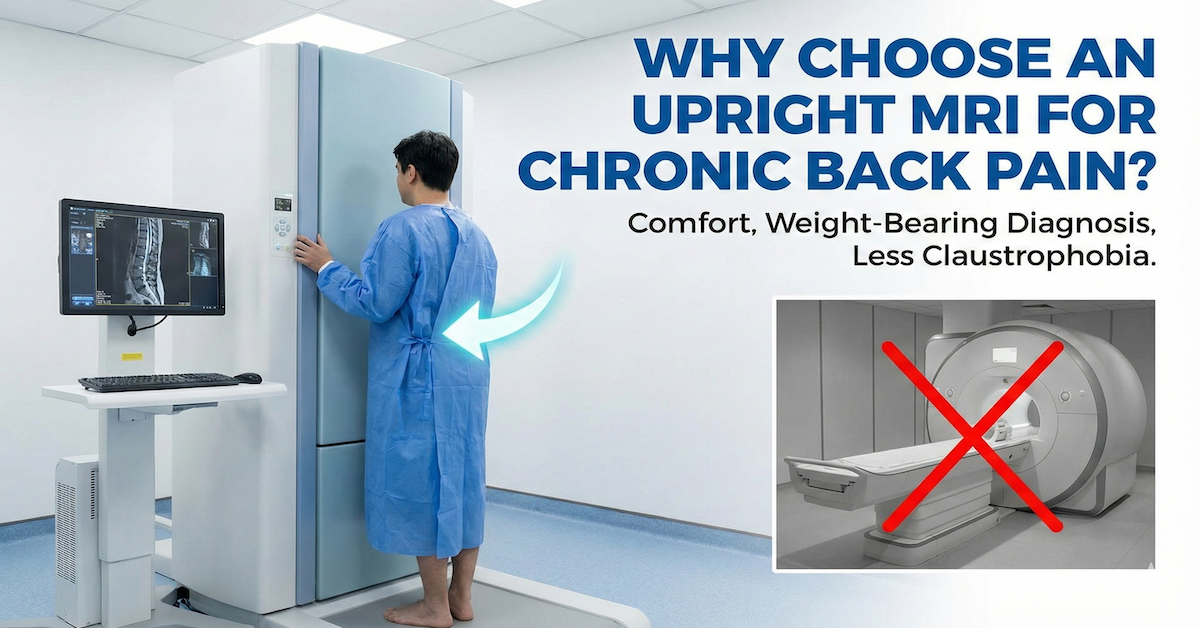
During the scan, you’ll lie on a table that slides into the MRI machine. For patients with claustrophobia, options like an open MRI or an upright MRI can make the experience more comfortable. The scan typically lasts 30–60 minutes, and you’ll need to stay still to ensure clear images. If dynamic imaging is needed, you may be asked to move your neck into specific positions.
After the scan, a radiologist will review the images and send a detailed report to your doctor. This collaborative effort ensures an accurate diagnosis and a clear path forward.
Limitations of MRI for CCI Diagnosis
While MRI is a powerful tool, it does have its limitations. Interpreting MRI images for CCI requires specialized knowledge, as the condition can involve very subtle changes. That’s why it’s essential to have an experienced radiologist analyze the scans.
Additionally, MRI isn’t suitable for everyone. Patients with certain types of metal implants or devices, such as pacemakers, may not be able to undergo an MRI. However, alternative imaging methods can be explored in such cases.
MRI also works best when combined with other diagnostic tools. Physical exams, patient history, and possibly complementary imaging like CT scans all contribute to a comprehensive understanding of the condition.
Treatment Implications of MRI Findings
Once an MRI confirms cranio-cervical instability, the results play a critical role in determining the next steps. For some patients, non-surgical approaches like physical therapy, bracing, or pain management may be sufficient. Others may require surgical intervention to stabilize the cranio-cervical junction.
MRI also helps monitor progress over time, allowing doctors to adjust treatments as needed. Follow-up scans can show whether ligaments are healing or if further intervention is necessary.
Conclusion
MRI scans have revolutionized the diagnosis of cranio-cervical instability, offering unmatched detail and accuracy. By providing clear images of ligaments, soft tissues, and alignment, MRI enables doctors to identify CCI with confidence and create effective treatment plans.
At Upright MRI of Deerfield, we specialize in advanced imaging that prioritizes both patient comfort and diagnostic precision. If you’re experiencing symptoms of cranio-cervical instability, we’re here to help you take the first step toward relief and recovery.
SHARE THIS POST:
Leave a Comment:

The World's Most Patient-Friendly MRI. A comfortable, stress-free, and completely reliable MRI scan. We offer patients an open, upright, standup MRI experience that helps those who are claustrophobic and stress being in a confined area. Upright MRI of Deerfield is recognized as the world leader in open MRI innovation,
Our Recent Post
READ PATIENT TESTIMONIALS
Upright MRI of Deerfield.
Susan D.,
Highland Park, 39
I am going to tell everyone about your office! This was a great experience after I panicked in other MRI machines and had to leave. Thank you so much.

Judith B.,
Milwaukee, 61
I suffer from vertigo and other MRIs do not work. This was wonderful…absolutely NO discomfort at all. The MRI was so fast…I wanted to stay and watch the movie! Mumtaz was great. His humor really put me at ease. I’ve already recommended Upright MRI to friends.

Delores P.,
Glencoe, 55
Everything is so nice and professional with your place. I have been there a couple of times. My husband and I would not go anywhere else.


