
457 Lake Cook Road (Deerfield Park Plaza)
Deerfield, IL 60015
Email Us:
Email us directly [+]
Fax: (847) 291-9362
How Can MRI Imaging Help Navigate Knee Health?
Knees play a vital role in mobility, and any discomfort or injury can significantly impact daily life. Whether it’s the result of a sports mishap, wear and tear over time, or an underlying condition, understanding the root cause of knee pain is essential. This is where MRI imaging steps in as a powerful diagnostic tool. It offers clarity and precision, helping doctors identify issues and tailor treatment plans effectively. Here’s a closer look at how MRI imaging can help you manage and improve your knee health.
What Is MRI Imaging and Why Is It Used for Knee Health?
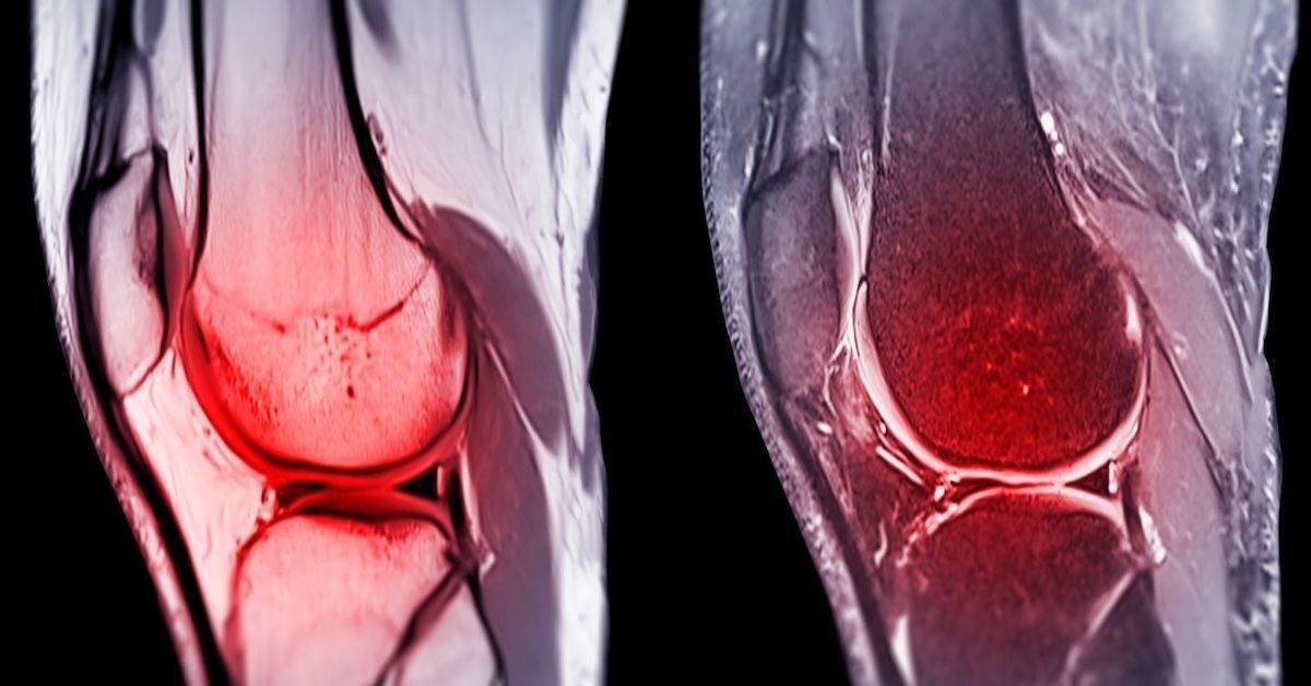
MRI, or Magnetic Resonance Imaging, is a non-invasive medical technology that uses magnetic fields and radio waves to create highly detailed images of the body’s internal structures. For knee health, MRI imaging is invaluable because it provides a comprehensive view of the soft tissues, cartilage, ligaments, tendons, and bones that make up the joint.
Unlike X-rays, which focus mainly on bones, MRIs give doctors the ability to see everything from torn ligaments to inflamed tissues. And because it doesn’t use radiation, MRI is a safe choice, especially for patients who may require repeated scans over time. It’s the gold standard for diagnosing knee injuries and conditions.
The Role of MRI in Diagnosing Knee Conditions
One of the main reasons doctors recommend an MRI for knee issues is its ability to reveal soft tissue injuries that aren’t visible on X-rays. For example, an MRI can identify torn ligaments like the ACL or MCL, common injuries among athletes. It’s also excellent at detecting meniscus tears, cartilage damage, or inflammation in the joint.
In cases of arthritis, MRIs go beyond simply confirming the presence of the condition. They provide detailed images of how the disease is affecting cartilage and surrounding structures, giving doctors a clearer understanding of its progression. Additionally, MRI imaging can spot infections or tumors, helping diagnose less common but serious issues.
How MRI Imaging Enhances Knee Injury Recovery
Getting an accurate diagnosis is the first step toward recovery, and MRI imaging ensures that doctors have all the information they need. By pinpointing the exact location and extent of an injury, MRIs help avoid misdiagnoses, which can lead to ineffective or unnecessary treatments.
Once the diagnosis is clear, MRI results guide the treatment plan. For minor injuries, this might mean physical therapy or medication. For more severe cases, like complete ligament tears, the scan can determine whether surgery is necessary. MRIs are also used during recovery to track progress and ensure that the treatment is working as expected.
Advantages of Using MRI for Knee Health
One of the standout benefits of MRI imaging is the level of detail it provides. Unlike other imaging techniques, an MRI captures the full picture of the knee, from ligaments and tendons to cartilage and bones. This makes it the preferred method for diagnosing complex injuries.
Another major advantage is its non-invasive nature. You won’t have to worry about surgery or long recovery times. It’s also completely painless and doesn’t involve radiation, making it a safer option, especially for those who might need multiple scans.
When Should You Consider an MRI for Your Knee?
If you’re experiencing persistent knee pain, swelling, or stiffness that hasn’t improved with rest or basic treatments, it might be time to consider an MRI. Injuries from sports or accidents often require an MRI to assess ligament or cartilage damage, especially if you’re struggling to bear weight on the affected knee.
MRIs are also helpful after surgery to evaluate healing and ensure that no complications arise during recovery. Even if your discomfort doesn’t seem injury-related, unexplained pain or limited range of motion could indicate underlying issues that an MRI can uncover.
Limitations of MRI Imaging for Knee Health
While MRIs are incredibly effective, they aren’t without limitations. For instance, the cost can be a concern for some patients, though insurance often covers the procedure when medically necessary. Accessibility can also be an issue in areas with limited imaging facilities.
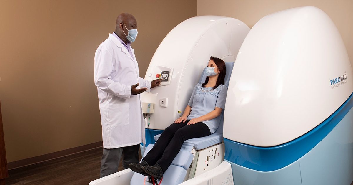
Claustrophobia is another factor, as traditional MRI machines involve lying in an enclosed space. Fortunately, open or upright MRI options can make the experience more comfortable for patients with anxiety. It’s also worth noting that MRIs work best when combined with a physical exam and other diagnostic tools, as they’re not a standalone solution.
Preparing for Your MRI Knee Scan
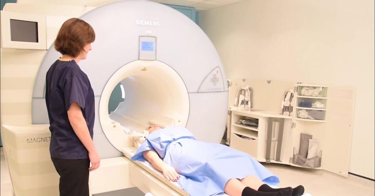
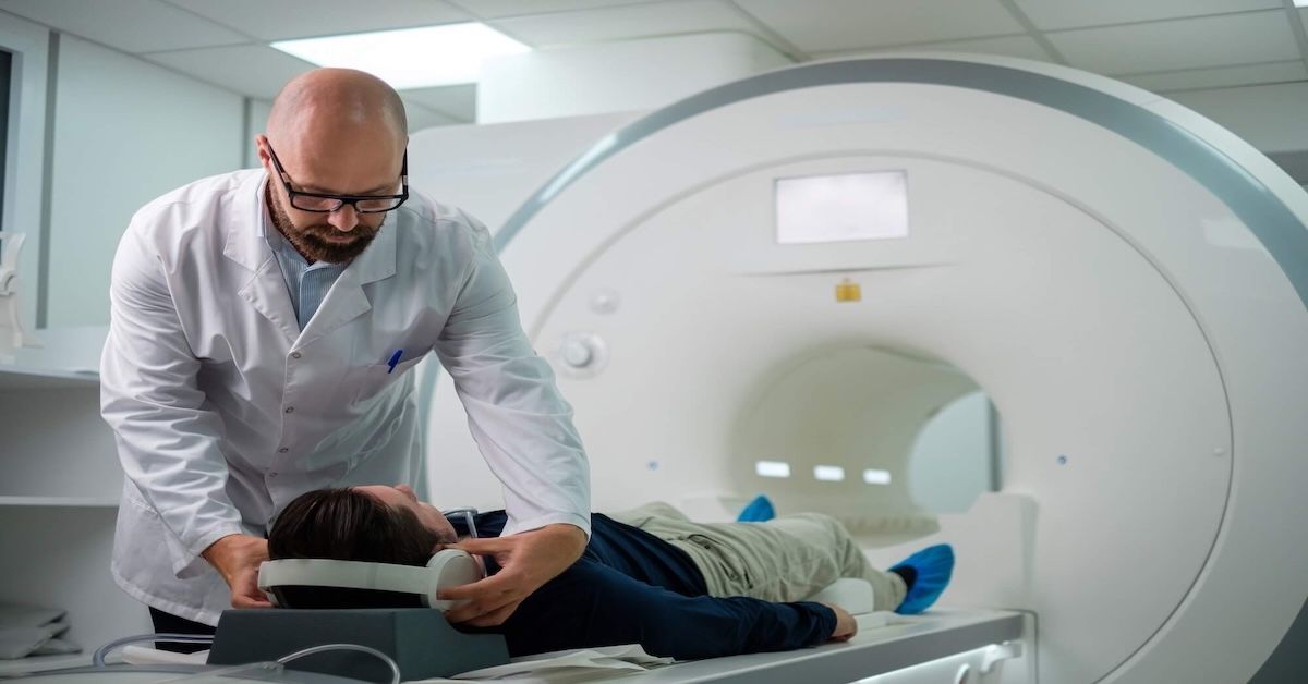
Preparing for an MRI is simple, but knowing what to expect can ease any nerves. You’ll likely need to remove metal objects like jewelry and wear comfortable clothing. Depending on the facility, you may be given a gown to change into.
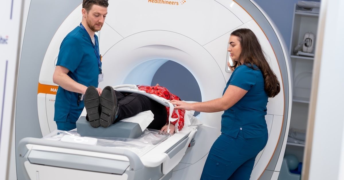
During the scan, you’ll lie on a table, and the machine will create detailed images of your knee. The process is painless, but you’ll hear loud, rhythmic sounds as the machine works. Most scans take about 30–45 minutes. Afterward, a radiologist will review the images and share the results with your doctor.
Conclusion
MRI imaging plays a crucial role in maintaining and improving knee health. Its ability to provide detailed, accurate insights makes it an essential tool for diagnosing injuries, guiding treatments, and monitoring recovery. Whether you’re dealing with an injury, chronic pain, or an unexplained issue, an MRI can offer the clarity you need to take the next step toward recovery.
At Upright MRI of Deerfield, we specialize in providing comfortable, high-quality imaging services tailored to your needs. If you’re experiencing knee pain or need a reliable diagnosis, our team is here to help. Schedule your scan today and take the first step toward better knee health.
SHARE THIS POST:
Leave a Comment:
The World's Most Patient-Friendly MRI. A comfortable, stress-free, and completely reliable MRI scan. We offer patients an open, upright, standup MRI experience that helps those who are claustrophobic and stress being in a confined area. Upright MRI of Deerfield is recognized as the world leader in open MRI innovation,
Our Recent Post


READ PATIENT TESTIMONIALS
Upright MRI of Deerfield.
Susan D.,
Highland Park, 39
I am going to tell everyone about your office! This was a great experience after I panicked in other MRI machines and had to leave. Thank you so much.

Judith B.,
Milwaukee, 61
I suffer from vertigo and other MRIs do not work. This was wonderful…absolutely NO discomfort at all. The MRI was so fast…I wanted to stay and watch the movie! Mumtaz was great. His humor really put me at ease. I’ve already recommended Upright MRI to friends.
Delores P.,
Glencoe, 55
Everything is so nice and professional with your place. I have been there a couple of times. My husband and I would not go anywhere else.

Follow UpRight MRI of Deerfield on Facebook
To see our latest news, updates or to get to know us more, we welcome you to follow along our journey in Facebook.


CONTACT DETAILS
Phone: (847) 291-9321
Address: 457 Lake Cook Road (Deerfield Park Plaza) Deerfield, IL 60015
Email: info@uprightmrideerfield.com
Business Hours
- Mon - Thu
- -
- Friday
- -
- Saturday
- -
- Sunday
- Closed