
457 Lake Cook Road (Deerfield Park Plaza)
Deerfield, IL 60015
Email Us:
Email us directly [+]
Fax: (847) 291-9362
How Does MRI Aid in Precision Imaging for Cranio-Cervical Instability?
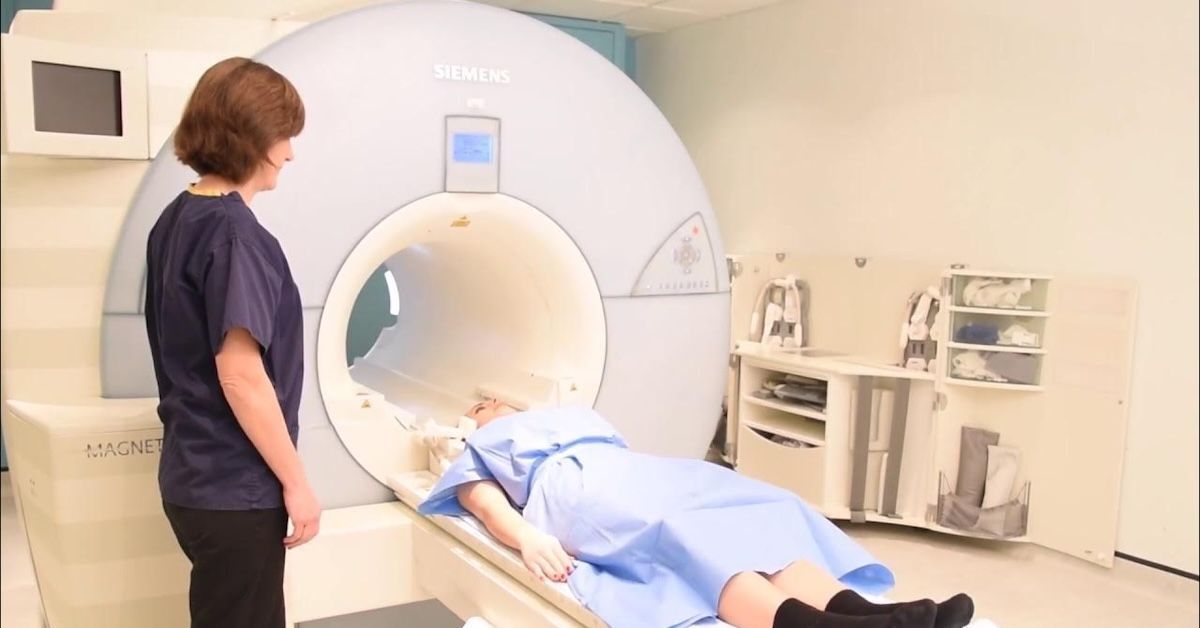
Cranio-cervical instability (CCI) is a complex condition that affects the joints where the skull and spine meet. When these structures become unstable, it can lead to a range of symptoms from chronic neck pain and headaches to dizziness and even neurological problems. Diagnosing CCI can be challenging, but advances in imaging technology, particularly MRI (Magnetic Resonance Imaging)—are helping doctors pinpoint the underlying issues more accurately than ever.
MRI offers a non-invasive way to see deep inside the body, providing detailed images of soft tissues, bones, and nerves. This allows for precise diagnostics, especially when other methods like X-rays fall short. In this article, we’ll explore how MRI helps in diagnosing CCI, what it can reveal, and why it’s considered the gold standard for this condition.

Understanding Cranio-Cervical Instability (CCI)
What is Cranio-Cervical Instability?
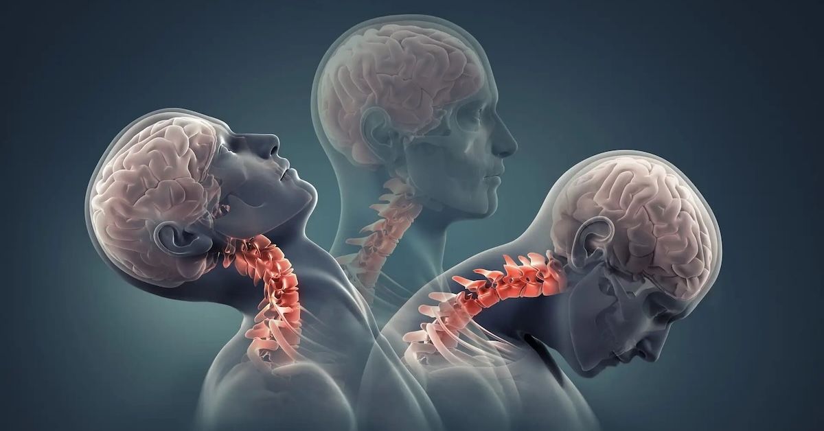
Cranio-cervical instability occurs when the ligaments and structures
connecting the skull and cervical spine are damaged or weakened. This instability allows for excessive movement between the head and spine, leading to a wide variety of symptoms. People with CCI might experience headaches, chronic neck pain, dizziness, balance issues, and even vision problems. The condition can be caused by trauma, such as whiplash or sports injuries, or by genetic disorders like Ehlers-Danlos syndrome, which affects connective tissue.
Why Accurate Diagnosis is Critical
The symptoms of CCI can overlap with those of other conditions, making diagnosis tricky. That’s why precise imaging is crucial—it allows doctors to see exactly what’s happening in the cranio-cervical junction. Accurate diagnosis through MRI enables targeted treatment, whether it’s physical therapy or, in severe cases, surgery. Without proper imaging, the root cause of the symptoms may go undetected, leading to ineffective treatments.
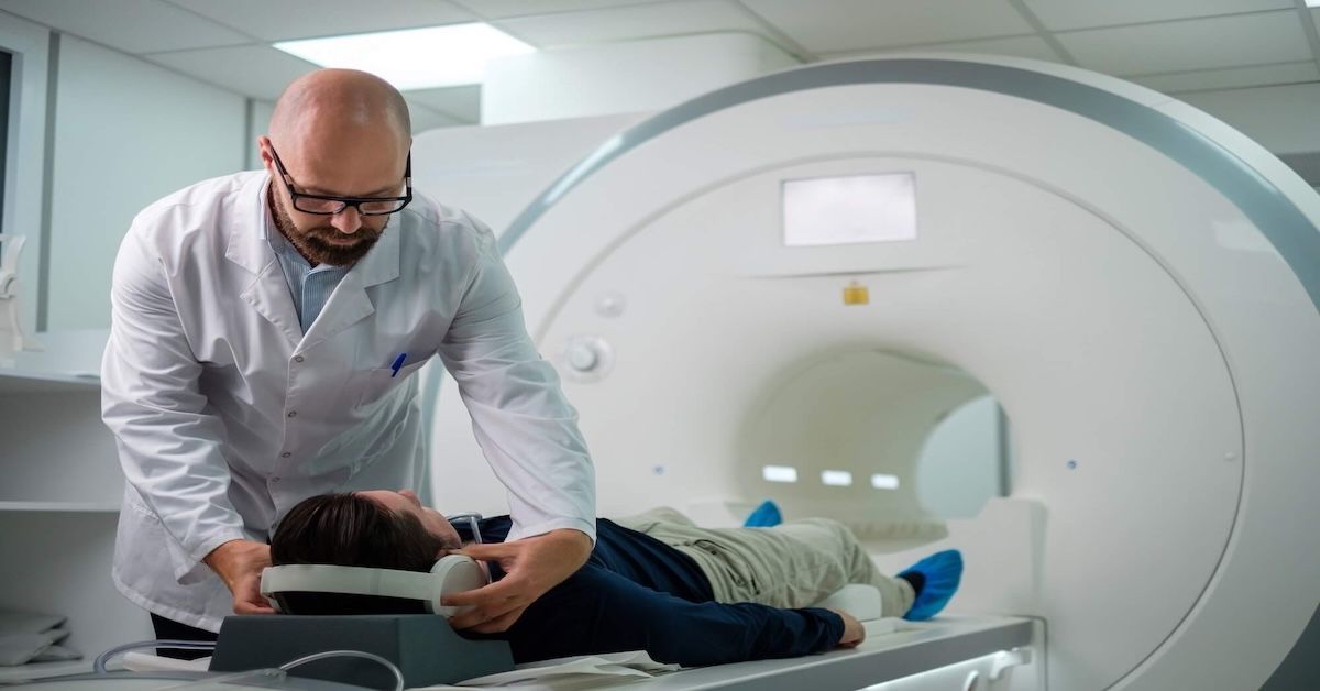
How MRI Works for Diagnosing CCI
MRI Technology and Its Unique Advantages
MRI uses powerful magnets and radio waves to create highly detailed images of the body’s internal structures, including bones, ligaments, and soft tissues. What sets MRI apart from other
imaging techniques like X-rays or CT scans is its ability to visualize soft tissues with exceptional clarity. This is particularly important for diagnosing CCI, where the condition often affects ligaments that are not visible on X-rays.
Why MRI is Ideal for CCI
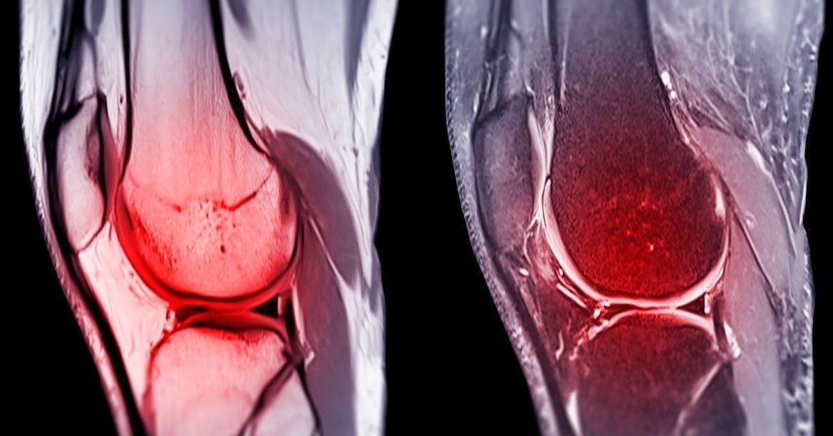
Because cranio-cervical instability often involves damage to the ligaments and soft tissues that support the skull and spine, MRI is the best imaging option. It offers clear images of the alar and transverse ligaments, two critical structures that help maintain stability in the cranio-cervical junction. MRI can also show how these ligaments are interacting with surrounding tissues, including the spinal cord and nerve roots, giving doctors a full picture of the condition.
Key Features MRI Can Identify in CCI Patients
Assessing Ligament Laxity or Rupture
One of the primary uses of MRI in diagnosing CCI is evaluating the health of the ligaments that support the cranio-cervical junction. Ligament laxity (weakening or loosening) or rupture (complete tearing) are common causes of instability. With MRI, doctors can see the exact condition of these ligaments, allowing them to determine whether non-surgical treatments like physical therapy might help or if surgery is required to stabilize the area.
Cervical Spine Alignment and Bone Structure
While CCI primarily affects the ligaments, bone structure plays a role as well. Misalignment of the vertebrae can contribute to the condition, and MRI provides detailed images of both bones and soft tissues. This allows doctors to assess whether misalignment or bone degeneration is worsening the instability. MRI can also show whether any small fractures are present, which might not be visible on a traditional X-ray.
Nerve and Spinal Cord Compression
One of the most serious complications of cranio-cervical instability is nerve compression. If the cranio-cervical junction is unstable, it can put pressure on the nerves or spinal cord, leading to neurological symptoms like tingling, numbness, weakness, or even cognitive difficulties. MRI can help doctors see if there is any nerve or spinal cord compression and gauge how severe it is, helping to plan the most appropriate treatment to prevent further damage.
MRI vs. Other Imaging Techniques for CCI
MRI vs. X-rays and CT Scans
While X-rays and CT scans are useful for looking at bone structure, they fall short when it comes to soft tissue imaging. X-rays can’t show ligaments or nerves, which are often the source of the problem in CCI. CT scans provide more detail than X-rays but still don’t offer the soft tissue clarity that MRI does. In short, MRI is far superior for diagnosing ligamentous injuries and other soft tissue abnormalities related to CCI.
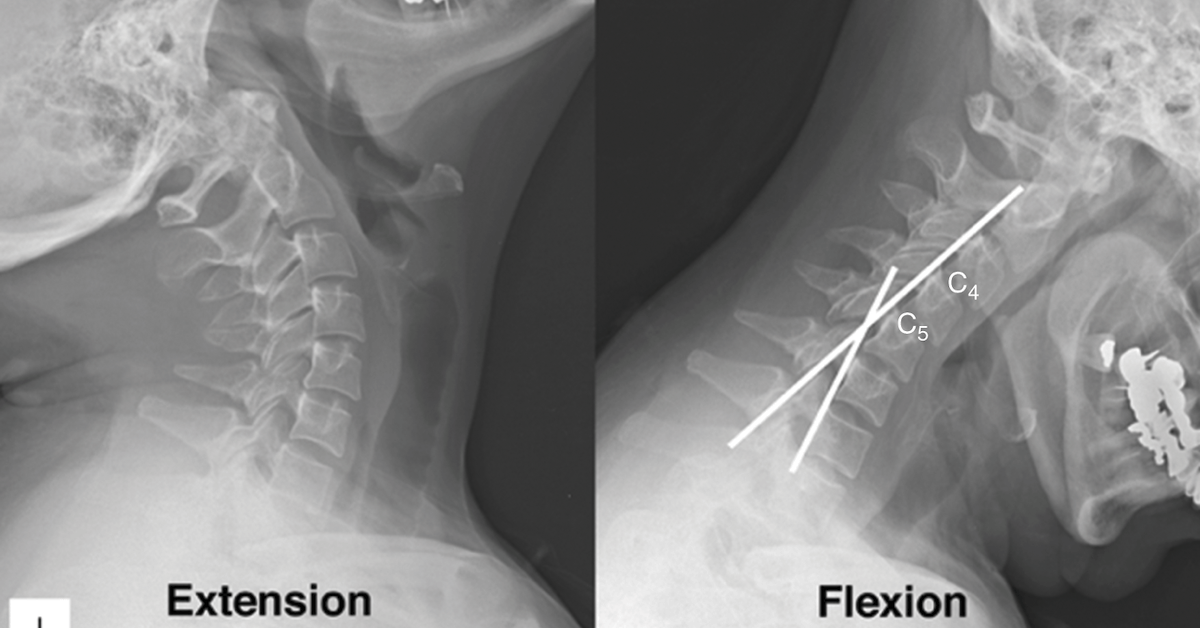
The Role of Functional MRI and Upright MRI in CCI
Functional MRI (fMRI) and Upright MRI offer even more insight for diagnosing CCI. Functional MRI captures images while the patient is in motion, helping doctors see how the
cranio-cervical junction behaves during specific movements. This is important for understanding how instability affects functionality in daily life. Upright MRI is another excellent tool for CCI diagnosis, as it allows doctors to capture images while the patient is sitting or standing, offering a better view of how gravity impacts cranio-cervical alignment.
When to Consider an MRI for CCI Symptoms
Persistent Neurological Symptoms
If you’ve been experiencing headaches, dizziness, or cognitive issues that don’t seem to go away, it might be time to consider an MRI. Neurological symptoms can often be caused by compression of the spinal cord or nerves in the cranio-cervical junction, and MRI is the best way to see what’s going on beneath the surface.
Unexplained Neck Pain and Restricted Movement
Chronic neck pain that hasn’t responded to treatment is another sign that an MRI may be necessary. Whether it’s pain when turning your head or discomfort while tilting it forward or back, an MRI can help identify whether
cranio-cervical instability is the culprit. This type of imaging is also useful for determining the extent of any ligament or bone damage that might be causing the pain.
Failure to Diagnose with Other Methods
If X-rays or CT scans have come back negative but your symptoms persist, an MRI can provide the clarity needed to make a proper diagnosis. MRI’s ability to see soft tissues and nerves makes it the most comprehensive imaging option for detecting CCI, especially in cases where other methods haven’t revealed the problem.
How MRI Results Guide Treatment for CCI
Tailoring Treatment Based on MRI Findings
Once an MRI has provided a clear diagnosis, doctors can tailor treatment to your specific condition. If the MRI shows ligament laxity but no nerve compression, non-invasive treatments like physical therapy or bracing may be recommended. However, if the scan reveals severe instability or nerve involvement, surgical options like fusion may be necessary to stabilize the area.
Monitoring Progress and Adjusting Treatment
MRI isn’t just useful for initial diagnosis—it can also help doctors monitor your progress over time. Follow-up MRIs can show whether treatments like physical therapy are improving stability or if additional interventions are needed. For patients who undergo surgery, MRI is often used to evaluate how well the cranio-cervical junction is healing and to ensure no new issues have developed.
Conclusion
MRI plays a critical role in diagnosing and managing cranio-cervical instability, offering unparalleled detail in imaging soft tissues, ligaments, and nerves. If you’re experiencing persistent symptoms like headaches, neck pain, or neurological issues, an MRI could provide the answers you need to start a proper treatment plan.
At Upright MRI of Deerfield, we specialize in offering comfortable, detailed MRI scans to help diagnose conditions like cranio-cervical instability. Our state-of-the-art equipment and expert staff ensure that you get the precise imaging needed to understand your condition and move forward with effective care. Contact us today to schedule your MRI and begin your path to relief.
SHARE THIS POST:
Leave a Comment:
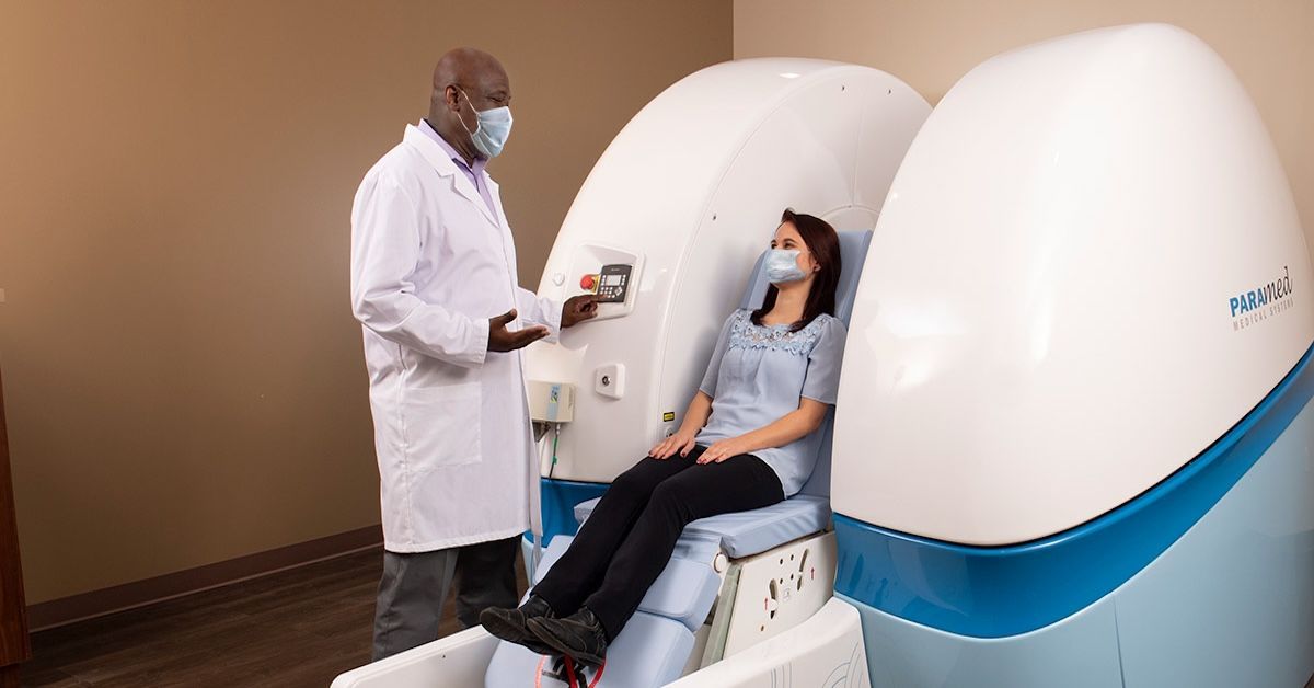
The World's Most Patient-Friendly MRI. A comfortable, stress-free, and completely reliable MRI scan. We offer patients an open, upright, standup MRI experience that helps those who are claustrophobic and stress being in a confined area. Upright MRI of Deerfield is recognized as the world leader in open MRI innovation,
Our Recent Post


READ PATIENT TESTIMONIALS
Upright MRI of Deerfield.
Susan D.,
Highland Park, 39
I am going to tell everyone about your office! This was a great experience after I panicked in other MRI machines and had to leave. Thank you so much.

Judith B.,
Milwaukee, 61
I suffer from vertigo and other MRIs do not work. This was wonderful…absolutely NO discomfort at all. The MRI was so fast…I wanted to stay and watch the movie! Mumtaz was great. His humor really put me at ease. I’ve already recommended Upright MRI to friends.
Delores P.,
Glencoe, 55
Everything is so nice and professional with your place. I have been there a couple of times. My husband and I would not go anywhere else.

Follow UpRight MRI of Deerfield on Facebook
To see our latest news, updates or to get to know us more, we welcome you to follow along our journey in Facebook.


CONTACT DETAILS
Phone: (847) 291-9321
Address: 457 Lake Cook Road (Deerfield Park Plaza) Deerfield, IL 60015
Email: info@uprightmrideerfield.com
Business Hours
- Mon - Thu
- -
- Friday
- -
- Saturday
- -
- Sunday
- Closed