
457 Lake Cook Road (Deerfield Park Plaza)
Deerfield, IL 60015
Email Us:
Email us directly [+]
Fax: (847) 291-9362
What Makes Upright MRI the Best Choice for Flexion and Extension Imaging?
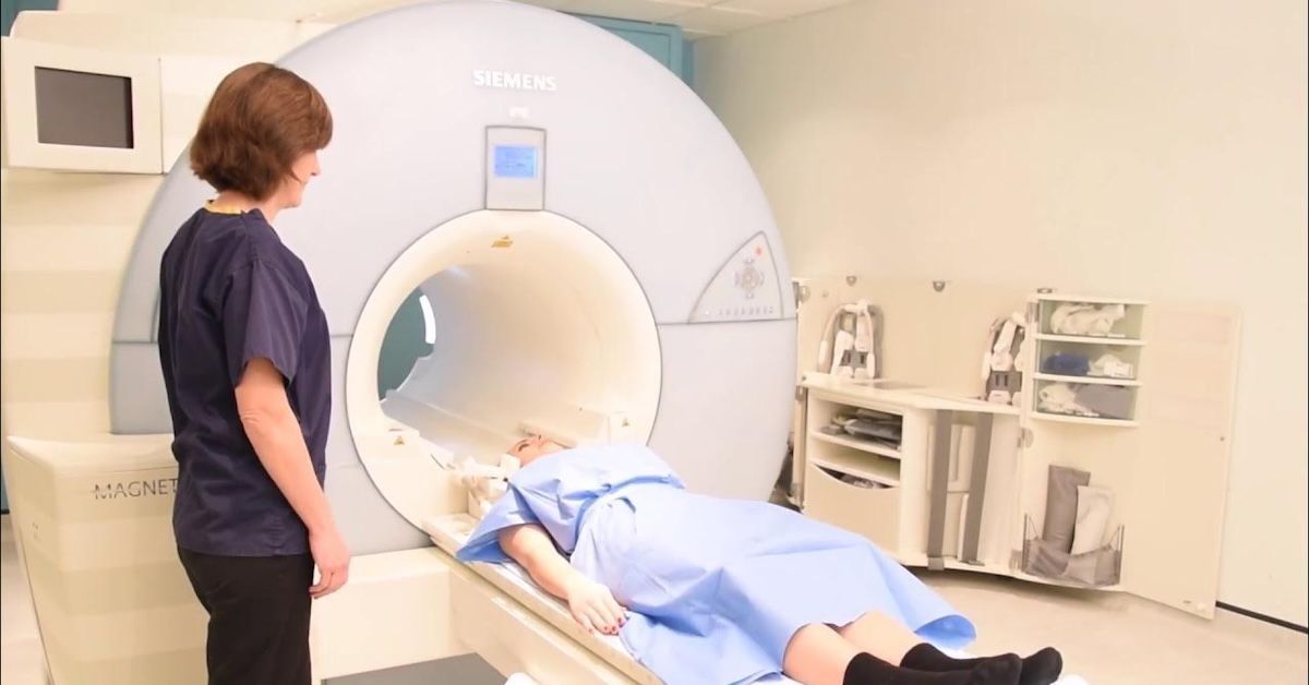
If you’ve been dealing with back or neck pain that seems to change depending on how you move, a regular MRI may not give you the answers you need. Traditional MRIs capture images while you’re lying flat, which doesn’t reflect how your spine behaves when you're sitting, standing, or bending. This is where flexion and extension imaging comes in. It allows doctors to see how your spine reacts to different movements, helping to detect issues that might not be visible in a standard scan.
Upright MRI is the best option for this type of imaging because it captures the spine in a natural, weight-bearing position. Unlike traditional MRIs, which only provide a still image, an upright MRI shows how the spine changes when bending forward or backward. This can reveal hidden spinal conditions that only appear when the body is in motion.
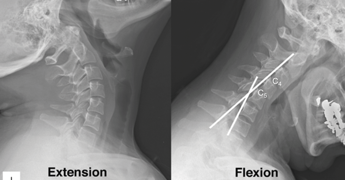
What Is Flexion and Extension Imaging and Why Does It Matter?
Flexion and extension imaging is a method used to evaluate spinal movement and stability. A regular MRI provides a single snapshot, but this type of scan looks at the spine in different postures to see how it responds to bending and stretching. For many patients, pain is not constant, it gets worse when they move in certain ways. This type of imaging helps pinpoint the exact movements that trigger discomfort.
By capturing images in multiple positions, doctors can diagnose conditions like spinal instability, herniated discs, and nerve compressions that may not be visible in a standard MRI. This is especially important for people who have had chronic back pain but no clear diagnosis from previous scans.
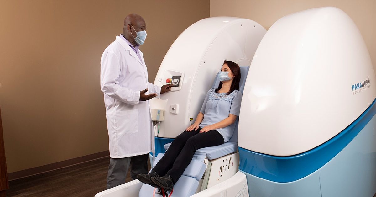
How Does Upright MRI Work for Flexion and Extension Scans?
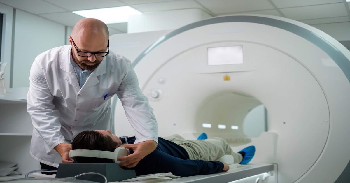
An upright MRI allows the patient to be scanned while sitting or standing, creating a much more accurate picture of how their spine functions in everyday life. This is different from traditional MRIs, which require the patient to lay motionless in a tube. Because the upright MRI is open, patients can be positioned in ways that mimic real-life movements.
During a flexion and extension scan, the patient bends forward and backward while the MRI captures images at each stage. This allows doctors to see how the spine moves, where it might be shifting incorrectly, and whether any discs or nerves are being compressed.
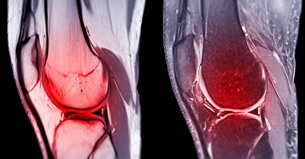
What Spinal Conditions Benefit from Flexion and Extension Imaging?
Many spinal problems are only noticeable when the body is in motion. A standard MRI may not catch them, but an upright MRI with flexion and extension imaging can.
Some conditions that are better diagnosed this way include:
- Herniated discs that bulge only when bending or standing
- Spinal stenosis, where the narrowing of the spine worsens in certain postures
- Spondylolisthesis, a condition where one vertebra slips over another when moving
- Cranio-cervical instability, which can lead to headaches, dizziness, and neck pain
- Whiplash injuries that affect soft tissues and spinal alignment
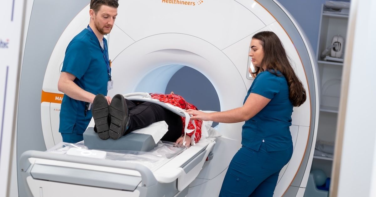
Why Traditional MRI Misses Flexion and Extension Issues
Regular MRIs have their place, but they don’t always tell the whole story. Since they are taken with the patient lying flat, they don’t account for how the spine reacts under real-life conditions. Many spinal problems only become evident when a person is standing, bending, or sitting, which means a standard MRI might come back normal even when a patient is in pain.
By using an upright MRI, doctors can see the spine under natural pressure. This is critical for diagnosing conditions that worsen when weight is applied, such as nerve compressions and shifting vertebrae. Without this kind of imaging, many patients might go undiagnosed or receive incorrect treatments based on incomplete information.
Advantages of Using Upright MRI for Flexion and Extension Scans
Upright MRI provides several benefits over traditional MRIs when it comes to flexion and extension imaging.
It allows for more accurate diagnoses because it captures images in positions where pain actually occurs. Instead of showing the spine at rest, it shows how it functions under normal conditions. This can be the key to identifying problems that have been missed before.
Patients also find upright MRI to be more comfortable. Since they can sit or stand during the scan, it eliminates the discomfort of lying in a confined tube for an extended period. For those with chronic pain or mobility issues, this can make the experience much easier.
For doctors and surgeons, upright MRI provides better insights for treatment planning. Whether a patient needs physical therapy, pain management, or surgery, having a detailed look at how their spine moves ensures that the chosen approach is the right one.
Who Should Consider an Upright MRI for Flexion and Extension Imaging?
This type of imaging is ideal for people experiencing pain that changes depending on movement or posture. If traditional MRIs have failed to explain your symptoms, a flexion and extension scan might provide the answers. It is particularly useful for those with chronic back or neck pain, previous injuries that never fully healed, or conditions that worsen when standing, sitting, or bending.
If you have been struggling with discomfort that comes and goes based on movement, this advanced imaging technique could be what finally leads to a proper diagnosis.
Conclusion
Flexion and extension imaging plays a crucial role in diagnosing spinal conditions that standard MRIs often miss. By scanning the spine in different postures, upright MRI provides a more complete picture of what’s happening inside the body. It is a valuable tool for both doctors and patients who need clear answers about movement-related pain.
At Upright MRI of Deerfield, we specialize in providing advanced imaging solutions, including flexion and extension MRI scans. If you’re dealing with ongoing pain and need a more accurate diagnosis, contact us today to learn more or schedule an appointment.
SHARE THIS POST:
Leave a Comment:
The World's Most Patient-Friendly MRI. A comfortable, stress-free, and completely reliable MRI scan. We offer patients an open, upright, standup MRI experience that helps those who are claustrophobic and stress being in a confined area. Upright MRI of Deerfield is recognized as the world leader in open MRI innovation,
Our Recent Post


READ PATIENT TESTIMONIALS
Upright MRI of Deerfield.
Susan D.,
Highland Park, 39
I am going to tell everyone about your office! This was a great experience after I panicked in other MRI machines and had to leave. Thank you so much.

Judith B.,
Milwaukee, 61
I suffer from vertigo and other MRIs do not work. This was wonderful…absolutely NO discomfort at all. The MRI was so fast…I wanted to stay and watch the movie! Mumtaz was great. His humor really put me at ease. I’ve already recommended Upright MRI to friends.
Delores P.,
Glencoe, 55
Everything is so nice and professional with your place. I have been there a couple of times. My husband and I would not go anywhere else.

Follow UpRight MRI of Deerfield on Facebook
To see our latest news, updates or to get to know us more, we welcome you to follow along our journey in Facebook.


CONTACT DETAILS
Phone: (847) 291-9321
Address: 457 Lake Cook Road (Deerfield Park Plaza) Deerfield, IL 60015
Email: info@uprightmrideerfield.com
Business Hours
- Mon - Thu
- -
- Friday
- -
- Saturday
- -
- Sunday
- Closed