
457 Lake Cook Road (Deerfield Park Plaza)
Deerfield, IL 60015
Fax: (847) 291-9362
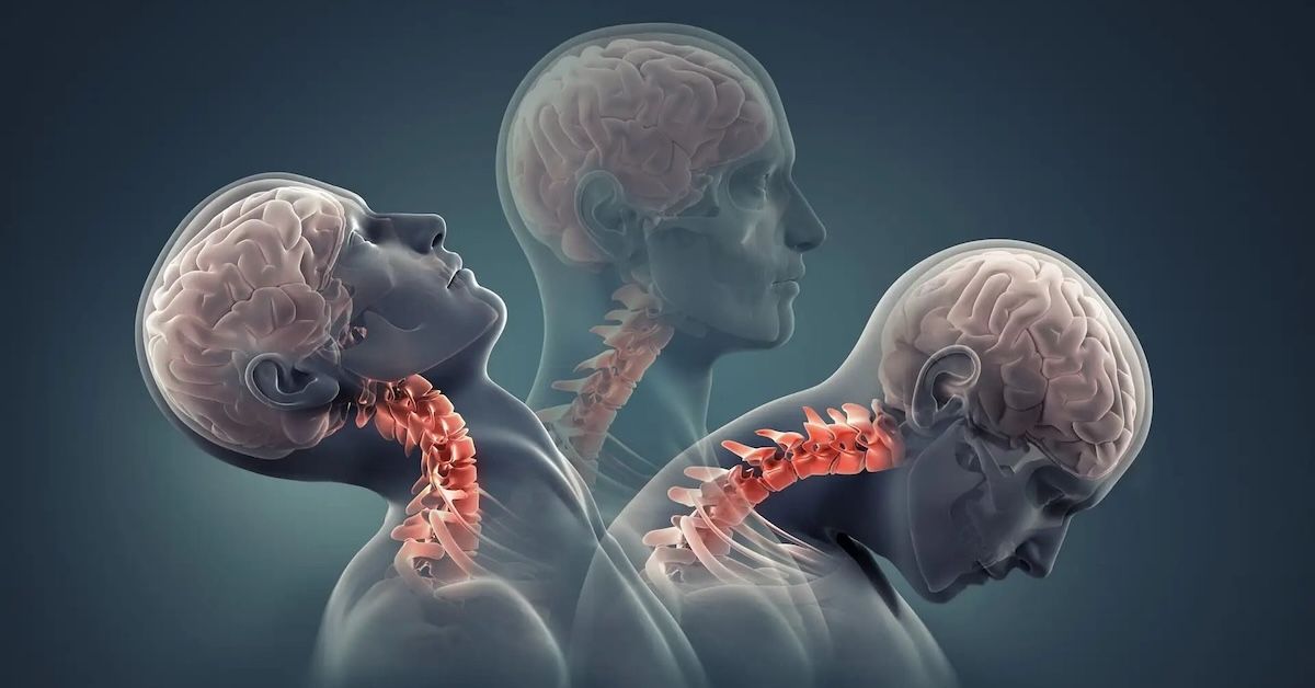
Why Are Traditional MRIs Missing Crucial Pathologies in the Spine?
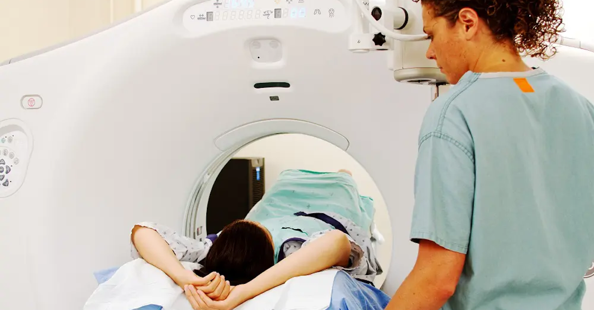
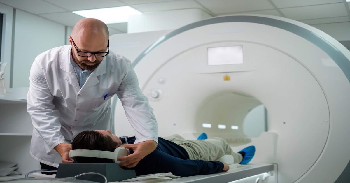
MRI scans are a cornerstone of spinal diagnostics. They provide valuable insights into the health of your spine, helping to identify issues like herniated discs, fractures, or degenerative conditions. However, despite being one of the most commonly used diagnostic tools, traditional MRIs are not always the most effective when it comes to detecting all spinal pathologies. There are several reasons why certain conditions might be missed, and this can have serious consequences for treatment and recovery. In this article, we’ll dive into why traditional MRIs can miss crucial spinal pathologies and explore the advancements in imaging that can fill these gaps.
Understanding Traditional MRI Technology
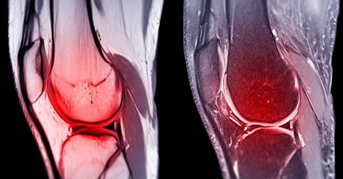
Traditional MRIs work by using magnetic fields and radio waves to produce images of the body’s internal structures. These machines are known for their ability to capture detailed pictures of soft tissues, like muscles, tendons, and the spinal cord. However, while MRIs excel at showing these soft tissues, they are not always as effective when it comes to highlighting smaller issues or subtle changes in the spine. The resolution of a traditional MRI may not be high enough to pick up early signs of disc degeneration, small fractures, or certain types of soft tissue injuries, which are key pathologies often seen in spinal conditions.
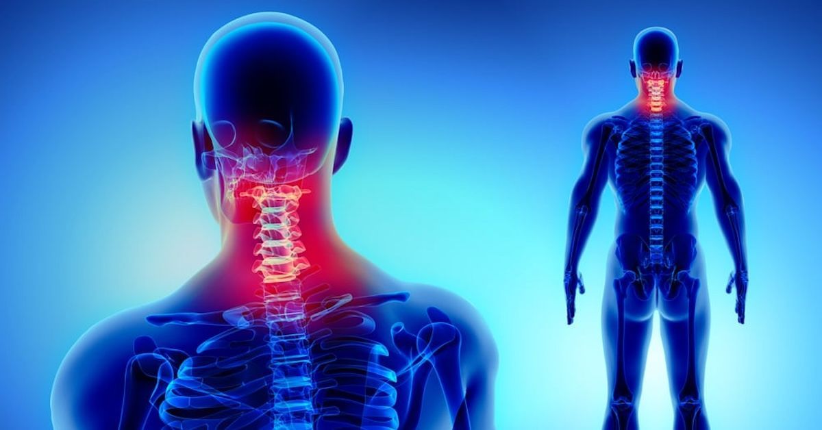
Common Pathologies Missed by Traditional MRIs
Despite their effectiveness, traditional MRIs sometimes miss critical spinal pathologies. Small disc herniations or subtle bulges, especially those that do not press significantly on nerve roots, might not show up on standard MRIs. In fact, many patients with symptoms like persistent back pain or tingling in the extremities may not have visible disc herniations on a traditional scan, leading to a misdiagnosis. Similarly, facet joint issues or ligament injuries can be missed, as MRIs don’t always provide clear images of these structures, especially in cases of mild damage. Another critical issue is hidden fractures or bone edema, which may be too small to detect with traditional imaging techniques, leading to undiagnosed issues that continue to affect the patient.
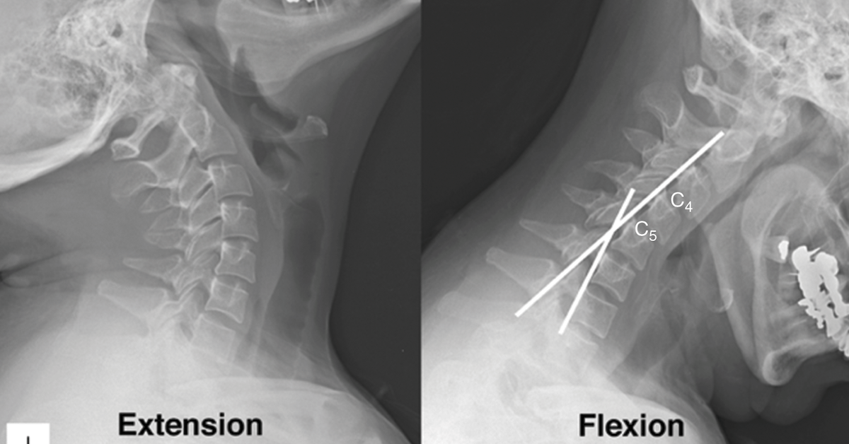
Factors That Affect MRI Accuracy in Detecting Spinal Pathologies
There are several factors that contribute to the inaccuracies of traditional MRIs. One of the most significant is the resolution of the images. If the scan’s resolution is too low, even small but crucial changes in the spine won’t appear clearly. Additionally, patient positioning during the scan can affect the clarity of the images. If a patient moves during the procedure or is not positioned in an optimal way, the resulting images can be blurry or distorted, which can lead to missed or incorrect diagnoses. Another challenge with traditional MRIs is their inability to effectively capture images of certain areas of the spine. Structures like the sacrum or deep regions of the cervical spine might not be visible enough on a standard MRI scan.
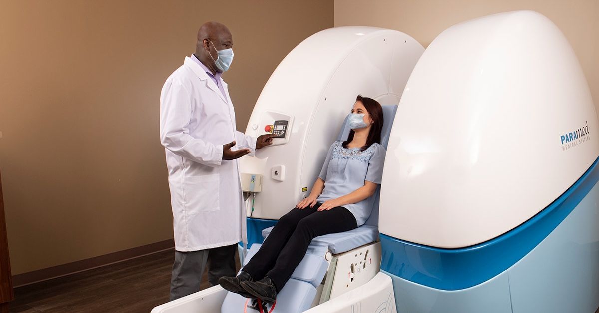
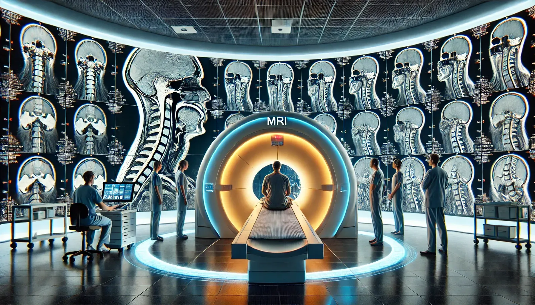
Alternative and Advanced Imaging Techniques
To overcome these limitations, healthcare providers are turning to more advanced imaging techniques. High-field MRIs, for example, provide more detailed and higher resolution images, allowing doctors to detect smaller issues that might be missed with traditional MRI machines. Another option is 3D MRI imaging, which provides a more comprehensive view of the spine, giving a clearer picture of both soft tissue and bone structures. For certain pathologies, CT scans or X-rays can also be used to offer more detailed images of the bones and vertebrae. Myelography, an imaging procedure where contrast dye is injected into the spinal canal, can help highlight abnormalities that might not be visible with conventional MRI scans. In some cases, a combination of these techniques can help provide a complete picture of the spine’s health.
Consequences of Missing Crucial Spinal Pathologies
When a spinal pathology is missed on an MRI, it can lead to serious consequences for the patient. A missed diagnosis often results in delayed treatments, which can allow the condition to worsen over time. For example, undiagnosed spinal fractures or herniations can lead to chronic pain, nerve damage, and other complications. The failure to detect conditions like spinal tumors or infections can be particularly dangerous, as these conditions may require urgent intervention to prevent further complications. Furthermore, missing subtle issues in the spine may result in inappropriate or ineffective treatments, leading to longer recovery times and unnecessary procedures.
The Future of Spinal Imaging
The future of spinal imaging looks promising, with many new technologies emerging to enhance the capabilities of MRIs. Newer MRI machines are providing higher resolution images and better contrast to detect even the smallest changes in spinal structures. Additionally, the integration of artificial intelligence (AI) and machine learning in imaging software is helping radiologists interpret MRIs more accurately, reducing the chances of missed diagnoses. AI algorithms are being trained to identify abnormalities that might otherwise go unnoticed, ensuring a more thorough and efficient review of MRI scans. These advancements will ultimately lead to earlier detection of spinal conditions, improving patient outcomes and reducing the need for invasive treatments.
Conclusion
Traditional MRIs are a valuable tool in spinal diagnostics, but they do have their limitations. Small herniations, subtle fractures, and certain types of soft tissue injuries can often be missed, leading to delayed or incorrect diagnoses. However, with the advent of advanced MRI technologies, improved imaging techniques, and AI-assisted diagnostics, these gaps are being addressed, allowing for more accurate and comprehensive evaluations of spinal health. At Upright MRI of Deerfield, we offer state-of-the-art imaging solutions designed to provide clear, detailed, and accurate results, ensuring that no pathology goes undetected. If you’re experiencing persistent spinal issues and feel that a traditional MRI might not be enough, reach out to us today to learn how our advanced imaging can help provide a better, more complete diagnosis.
SHARE THIS POST:
Leave a Comment:
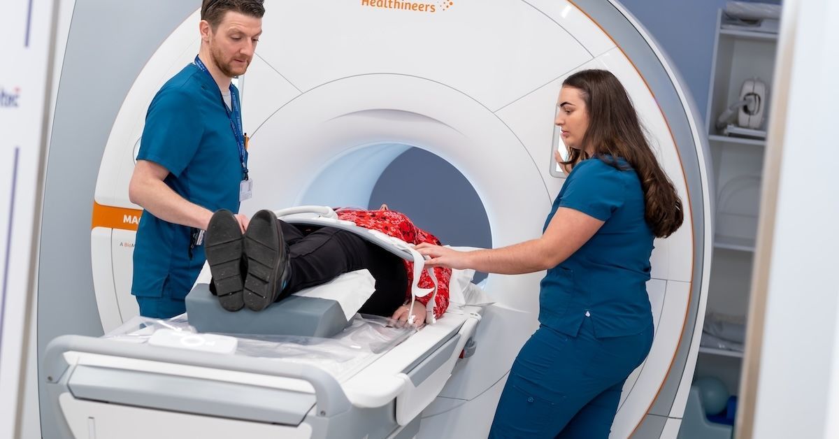
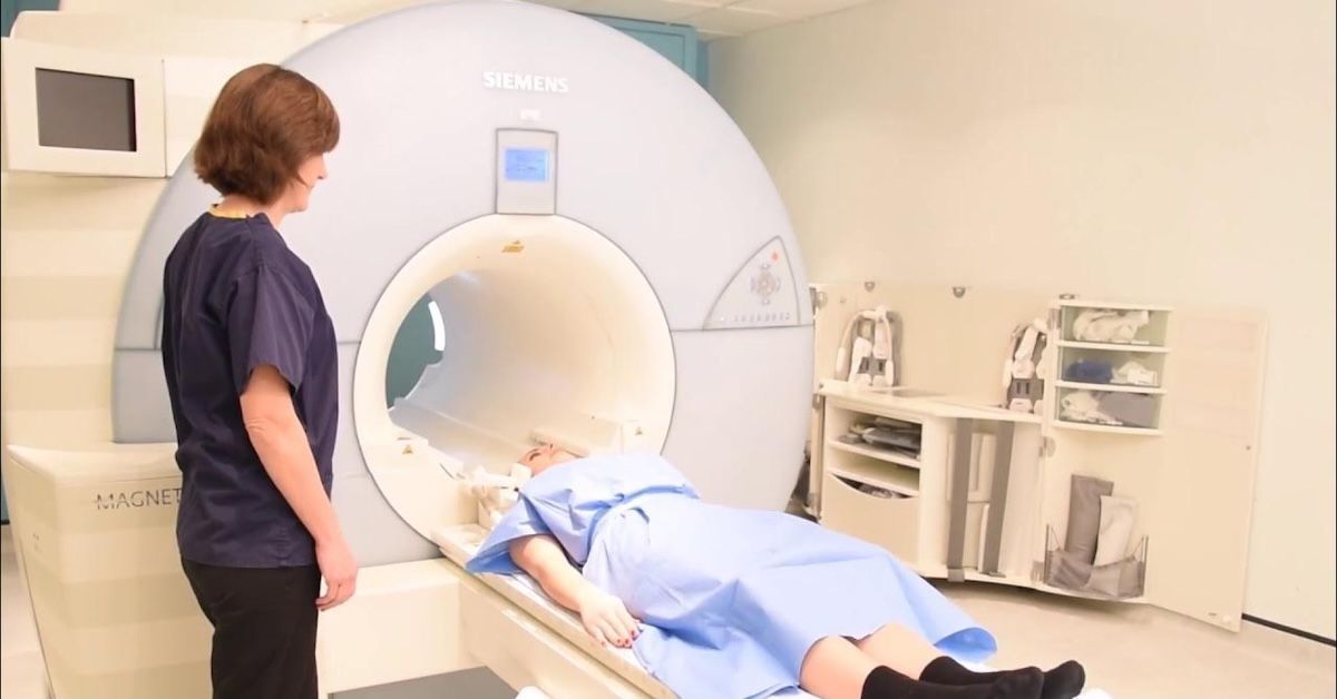
The World's Most Patient-Friendly MRI. A comfortable, stress-free, and completely reliable MRI scan. We offer patients an open, upright, standup MRI experience that helps those who are claustrophobic and stress being in a confined area. Upright MRI of Deerfield is recognized as the world leader in open MRI innovation,
Our Recent Post

READ PATIENT TESTIMONIALS
Upright MRI of Deerfield.
Susan D.,
Highland Park, 39
I am going to tell everyone about your office! This was a great experience after I panicked in other MRI machines and had to leave. Thank you so much.

Judith B.,
Milwaukee, 61
I suffer from vertigo and other MRIs do not work. This was wonderful…absolutely NO discomfort at all. The MRI was so fast…I wanted to stay and watch the movie! Mumtaz was great. His humor really put me at ease. I’ve already recommended Upright MRI to friends.
Delores P.,
Glencoe, 55
Everything is so nice and professional with your place. I have been there a couple of times. My husband and I would not go anywhere else.

Follow UpRight MRI of Deerfield on Facebook
To see our latest news, updates or to get to know us more, we welcome you to follow along our journey in Facebook.


CONTACT DETAILS
Phone: (847) 291-9321
Address: 457 Lake Cook Road (Deerfield Park Plaza) Deerfield, IL 60015
Business Hours
- Mon - Thu
- -
- Friday
- -
- Saturday
- -
- Sunday
- Closed